
Figures & data
Figure 1. Schematic representation of the formation of the growth plate during endochondral bone development. In the resting zone, chondrocytes have a round shape and they mainly express SOX9, Col2a1 and HIF-1α. In the proliferating layer, chondrocytes express SOX9, Acan, Col2a1, PTHrP, FGFR3; they assume a cuboidal shape, progressively pile up in columns. Columnar chondrocytes stop proliferating and they firstly become pre-hypertrophic cells, which express PTHrP receptor and Ihh. These cells terminally differentiate into hypertrophic cells, which express Col10a1 and VEGF; thus promoting angiogenesis. Lastly, hypertrophic cells are progressively replaced by osteoblasts expressing Col1a1 and osteopontin.
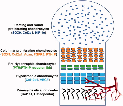
Figure 2. Schematic representation of EGF signalling pathway. The binding of EGF or EGF-like proteins induces oligomerization of EGFR (ErbB1) with ErbB2/c-neu, ErbB3, and ErbB4, and subsequent phosphorylation of the receptor, leading to the transduction of different pathways: PLC-γ1, PI-3 Kinase, GAP-MAP-raf kinase. These pathways are involved in several functions, such as cell proliferation, migration and survival.
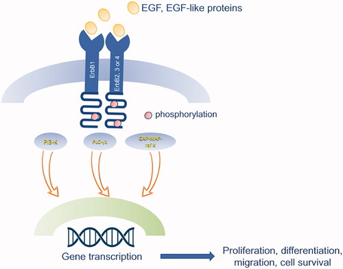
Figure 3. PRISMA flow diagram of the present systematic review.
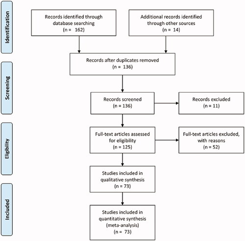
Table 1. EGF in bone metabolism database.
Table 2. EGF and endochondral ossification database.
Data availability statement
Data sharing is not applicable to this article as no new data were created or analysed in this study.