
Figures & data
Figure 1. Flow chart of this study.
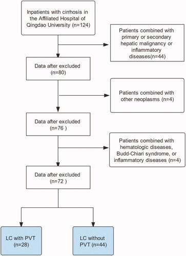
Table 1. Baseline demographics and clinical characteristics of the patients.
Figure 2. Receiver operating characteristic curve of a systemic immune-inflammation index (SII), neutrophil-to-lymphocyte ratio (NLR), monocyte-to-lymphocyte ratio (MLR), and platelet-to-lymphocyte ratio (PLR). The cut-off values for SII, NLR, MLR, and PLR were 268.9 (area under the curve, 0.612; p < .001), 3.14 (area under the curve, 0.596; p < .001), 0.37 (area under the curve, 0.662; p < .001), and 103.4 (area under the curve, 0.628; p < .001), respectively.
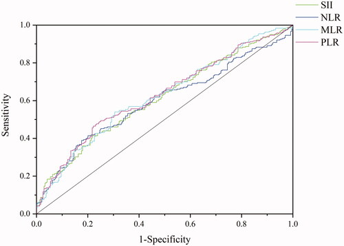
Table 2. Results of univariate analysis.
Table 3. Results of the multivariate logistic regression analysis.
Figure 3. Nomogram to predict the probability of portal vein thrombosis (PVT). To use this nomogram, the specific value for each patient should be located on each variable axis, and a line plotted upward to determine the points for each variable value. The sum of the points can be found on the “Total Points axis,” and a perpendicular line drawn downwards determines the risk of PVT.
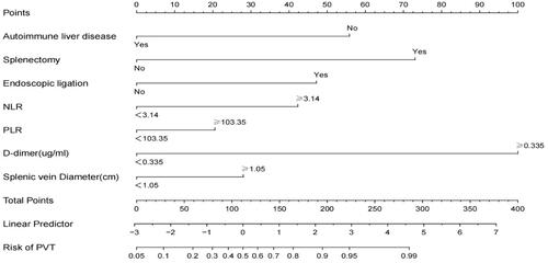
Figure 4. Receiver operating characteristic (ROC) curve of the model. The area under the ROC curve was 0.891 (95% confidence intervals 0.862–0.919).
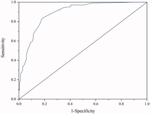
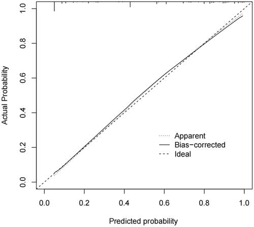
Figure 5. Calibration plots of the nomogram.
Data availability statement
The data that support the findings of this study are available from the author, upon reasonable request.