
Figures & data
Figure 1. Clinical manifestations of brittle nail syndrome. (A) Lamellar onychoschizia. (B) Onychorrhexis.
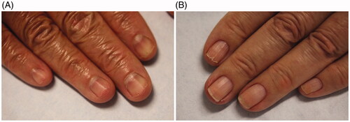
Table 1. Secondary causes of brittle nails.
Figure 2. Dermatophytoma (DOI: 10.1016/j.jaad.2018.03.062, Permission for reuse of this image has been obtained from the copyright holder (Elsevier) and applies to publications with the Creative Commons license).
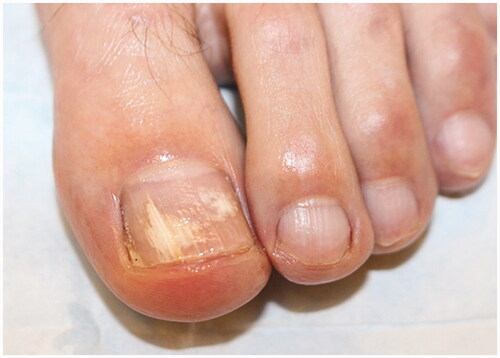
Figure 3. Clinical examination findings in onychomycosis. (A) Tinea pedis, scale on the plantar feet and web spaces. (B) Subungual hyperkeratosis and onycholysis of the right great toenail. (C) Yellow discolouration and onycholysis of the left great toenail. (D) Subungual hyperkeratosis and onycholysis in multiple toenails. (E) Severe onychodystrophy in multiple toenails. (DOI: 10.1016/j.jaad.2018.03.062, Permission for reuse of these images has been obtained from the copyright holder (Elsevier) and applies to publications with the Creative Commons license).
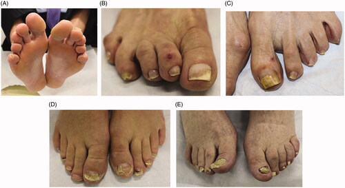
Table 2. Differential diagnoses for onychomycosis.
Figure 4. Nail pitting and onycholysis in right fingernails.
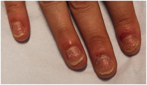
Table 3. Nail matrix and nail bed features of NP.
Figure 5. Subungual haematoma with onychomadesis.
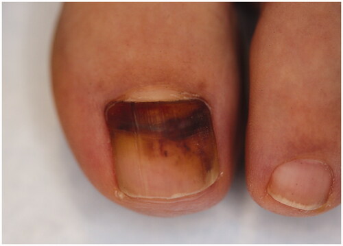
Figure 6. Clinical and dermoscopic findings of benign LM. (A) Ethnic melanonychia. On dermoscopy, grey background with multiple thin grey lines. (B) Melanocytic activation secondary to onychomycosis of the right thumbnail. (C) Junctional naevus. (DOI: 10.1016/j.jaad.2018.08.033, Permission for reuse of these images has been obtained from the copyright holder (Elsevier) and applies to publications with the Creative Commons license).
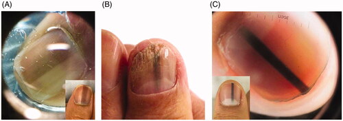
Figure 7. Subungual melanoma, 6 mm brown band. (DOI: 10.1016/j.jaad.2018.08.033, Permission for reuse of this image has been obtained from the copyright holder (Elsevier) and applies to publications with the Creative Commons license).
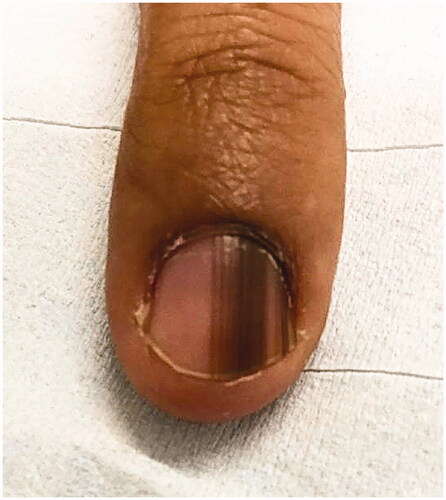
Figure 8. Dermoscopic findings of subungual melanoma. (A) Irregular colour, thickness and spacing without loss of parallelism. (B) Irregular colour, thickness and spacing with loss of parallelism. (DOI: 10.1016/j.jaad.2018.08.033, Permission for reuse of these images has been obtained from the copyright holder (Elsevier) and applies to publications with the Creative Commons license).
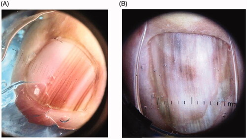
Figure 9. Systematic approach to LM.
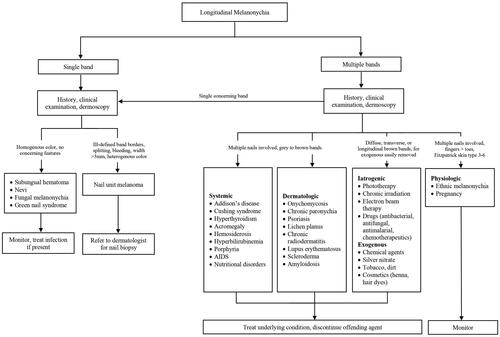
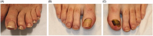
Figure 10. Clinical presentations of Beau’s lines, onychomadesis and retronychia. (A) Beau’s lines on the left toenails. (B) Onychomadesis of the left great toenail. (C) Retronychia of the right great toenail.
Data availability statement
Data sharing is not applicable to this article as no new data were created or analysed in this study.