
Figures & data
Figure 1. ASF1B is highly expressed in LGG tissues and is associated with poor prognosis of LGG patients. (A) GEPIA database showed that ASF1B was highly expressed in LGG patients. (B) GEPIA database showed that high ASF1B expression was significantly associated with low overall survival rate in LGG patients. (C) GEPIA database showed that high ASF1B expression was significantly associated with low disease-free survival rate of LGG patients. (D and E) RT-qPCR and Western blot were used to detect the expression of ASF1B in LGG cell lines. **p < 0.01, ***p < 0.001 vs NHA.
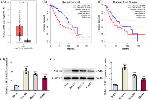
Table 1. Logistic regression analysis of ASF1B expression (univariate Cox analysis).
Table 2. Logistic regression analysis of ASF1B expression (multivariate Cox analysis).
Figure 2. Interference with ASF1B inhibits proliferation of LGG cells. (A and B) RT-qPCR and Western blot were used to detect the expression of ASF1B after transfection. C.CCK-8 detected the cell viability. (D and E) Colony formation assay was used to detect the cell proliferation. (F) Western blot was used to detect the expression of PCNA and Ki67. ***p < 0.001 vs sh-NC.
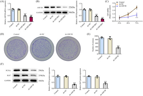
Figure 3. Interference with ASF1B induces cell cycle arrest and apoptosis in LGG. (A and B) Cell cycle was detected by flow cytometry. (C and D) TUNEL assay detected cell apoptosis. (E) Western blot was used to detect the expression of cell cycle and apoptosis-related proteins. ***p < 0.001 vs sh-NC.
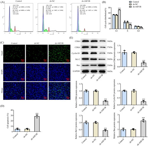
Figure 4. Interference with ASF1B inhibits invasion and migration of LGG cells. (A and B) Wound healing was used to detect the cell migration. (C and D) Transwell was used to detect the cell invasion. (E) Western blot was used to detect the expression of cell migration and invasion-related proteins. *p < 0.05, ***p < 0.001 vs sh-NC.
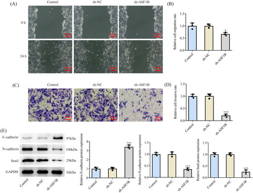
Figure 5. TLK1 interacts with ASF1B. STRING (A) and GeneMANIA (B) databases uncovered potential interactions between ASF1B and TLK1. (C and D) RT-qPCR and Western blot were used to detect the expression of TLK1 in LGG cells. ***p < 0.001 vs NHA. (E and F) The targeted binding of ASF1B and TLK1 was verified by ChIP assay. ***p < 0.001 vs IgG. (G and H) RT-qPCR and Western blot were used to detect the expression of TLK1 after depletion of ASF1B. ***p < 0.001 vs sh-NC.
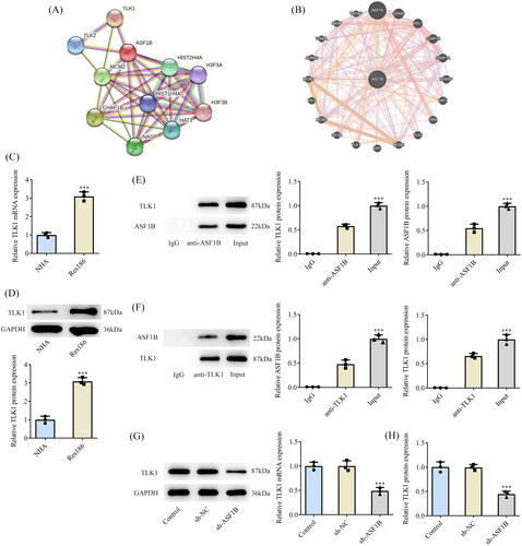
Figure 6. Upregulation of TLK1 partially reverses the effect of ASF1B interference on cell proliferation. (A and B) RT-qPCR and Western blot were used to detect the expression of TLK1 after transfection. ***p < 0.001 vs Ov-NC. (C) CCK-8 detected the cell viability. (D and E) Colony formation assay was used to detect the cell proliferation. F. Western blot was used to detect the expression of PCNA and Ki67. *p < 0.05, ***p < 0.001 vs Control; #p < 0.05, ##p < 0.01 vs sh-ASF1B + Ov-NC.
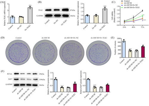
Figure 7. Upregulation of TLK1 partially reverses the effects of ASF1B interference on cell apoptosis. (A and B) Cell cycle was detected by flow cytometry. (C and D) TUNEL assay detected cell apoptosis. E and F. Western blot was used to detect the expression of cell cycle and apoptosis-related proteins. ***p < 0.001 vs Control; #p < 0.05, ###p < 0.001 vs sh-ASF1B + Ov-NC.
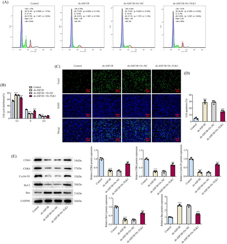
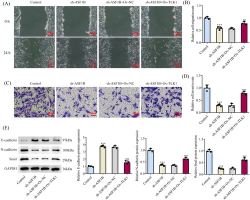
Figure 8. Upregulation of TLK1 partially reverses the effects of ASF1B interference on cell migration and invasion. (A and B) Wound healing was used to detect the cell migration. (C and D) Transwell was used to detect the cell invasion. (E) Western blot was used to detect the expression of migration and invasion-related proteins. ***p < 0.001 vs Control; ##p < 0.01, ###p < 0.001 vs sh-ASF1B + Ov-NC.
Data availability statement
The analysed data sets generated during the present study are available from the corresponding author on reasonable request.