
Figures & data
Figure 1. Timeline of experiment procedures.
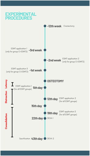
Figure 2. Unfocussed applicator of ESWT and shock wave application to distraction area.
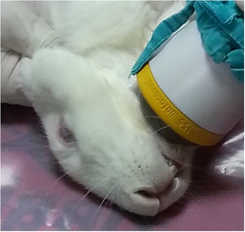
Figure 3. The custom-made titanium distractor and osteotomy line is posterior to the mental foramen. Distractor was positioned parallel to the lower border of the mandible.
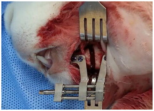
Figure 4. (A) DEXA. (B,C) Three-dimensional (CT) images showed healthly distracted bone and unilateral crossbite.
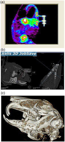
Table 1. BMD data obtained from DEXA examinations at 1st and 4th week of consolidation (g/cm2).
Figure 5. Histological image showing connective tissue areas of (A,D–F). The connective tissue areas are marked with red star. New bone areas have been shown in (G,J–L) images. New bone areas are marked with a white star. New vessel areas have been shown in (M,P,R,S) images. New vessel areas are marked with white (→). (original magnification ×5, hematoxylin-eosin).
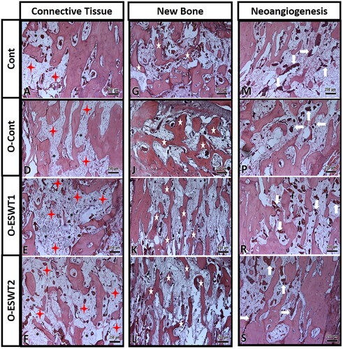
Table 2. Tissue volumes obtained from the stereological examination (mm3).
Data availability statement
The data that support the findings of this study are available from the corresponding author, [Dr.Enes Özkan], upon reasonable request.