
Figures & data
Table 1. Clinical characteristics.
Figure 1. The mass on the infant’s left upper limb was found immediately after birth.
a: Lesions located on the left upper limb in a 24-day-old boy, with the surface swollen and purple. b: The angiography of the left brachial artery showed that the feeding arteries of the lesion came from multiple brachial artery branches. c: The swelling was alleviated, tumour size was reduced and skin colour got recovered 1 month after TACE. d: The tumour was completely absorbed and the surface skin was flabby 6 months after TACE.
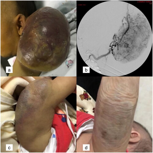
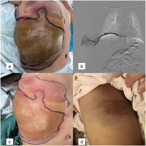
Figure 2. The lesion on the right abdominal wall of a 4-month-old boy.
a: Lesions in the right abdominal wall. The inner ring showed the range of lesions in the first day of hospitalization, and the outer ring showed the range of lesions in the 2nd day of hospitalization. b: One of feeding vessels of the lesion, tortuous intercostal artery. c: The swelling was alleviated 1 week after TACE. d: The tumour was completely absorbed and a little pigment deposition was remained 6 months after TACE.
Data availability statement
The datasets used and/or analyzed during the current study available from the corresponding author on reasonable request.