
Figures & data
Table 1. General information of the patients (mean ± SD).
Figure 1. The 3D model of the right adrenal gland of a 40-year-old male patient. (A) Right anterior view. (B) Left posterior view. (C) Inferior posterior view.
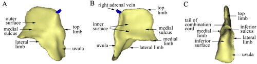
Figure 2. The 3D structure of adrenal glands. (A) The overall shape of the right adrenal gland with a deeper inferior sulcus looked like the ‘llama hat’ in a 32-year-old male patient. (B) The overall shape of the right adrenal gland was similar to the triangular leaf of a pitaya in a 62-year-old female patient.
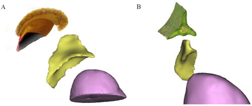
Figure 3. Effect of fusion of the adrenal venographic image and the 3D model. (A) An anteroposterior fusion image of a 50-year-old male patient. The right central vein was in the axial direction, and its image was a point at the catheter tip. The venographic image conformed to the ‘starburst type‘. Veins in the ‘starburst type’ were distributed in the upper part of the 3D model, while the uvula veins in the lower part of the 3D model were not shown. (B) An anteroposterior fusion image of a 34-year-old male patient. The right central vein was in the oblique axial direction and gradually shown, forming a ‘solid triangle type’ with its branches. The three limb veins were distributed in three limbs, referring to the central vein as the centre. No visible veins were shown in the uvula. (C) An anteroposterior fusion image of a 52-year-old female patient. The right central vein was further shown and communicated with renal capsular veins, forming a ‘hollow triangle type‘. The brush vein shown could be a signature vein of adrenal veins. No visible veins were shown in the uvula.
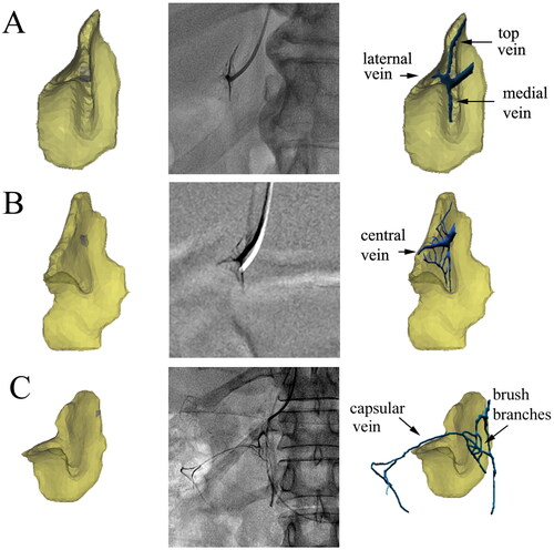
Figure 4. Effect of fusion of the irregular type and 3D model. (A) Adrenal fusion image of a 52-year-old female patient. The distribution of the adrenal veins was disordered, forming an ‘irregular type‘. Brush veins and central veins were shown at the catheter tip. (B) In the same patient, because of the catheter tip bending to the medial gland, more contrast agents entered the uvula vein.
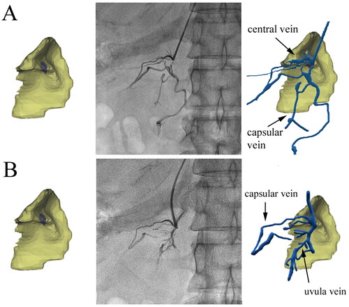
Table 2. Morphology classification of right adrenal venography (n = 41).
Figure 5. Effect of fusion of the trunk branch type and 3D model. (A) An anteroposterior fusion image of a 37-year-old female patient. The course of the central vein was close to the horizontal direction, and the uvula veins extended downward, with multiple branches distributed in the uvula. (B) Adrenal fusion image at 30 degrees right anterior oblique of a 47-year-old male patient. The angle between the uvula and central veins was close to 90 degrees. The distribution of branches of the uvula veins was similar to leaf texture.
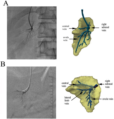
Figure 6. The signature veins of the right adrenal gland. (A) A 27-year-old male patient’s brush vein (long white arrow) almost vertically drained into the central vein (black arrow). The renal capsular vein (short white arrow) communicated with the brush vein expanded laterally. (B) In the ‘spider type’ of a 46-year-old male patient, the central vein (short black arrow) and the uvula vein (long black arrow) can be distinguished, but the brush vein was not shown. It could be seen that the renal capsular veins (white arrows), like soft vines, drained into nearly the outlet of the central vein. The overall shape of these renal capsular veins was similar to the spider’s foot. (C) A 42-year-old male patient’s venographic image showed relatively complete adrenal veins: the central vein (short black arrow), the uvula vein and its branch veins (long black arrow), the lateral limb vein (short white arrow), and the renal capsular vein (long white arrow) communicated with the distal end of the central vein. (D) The ‘solid triangle type’ of a 57-year-old female patient included the solid upper triangle venous enhancement area (short white arrow) and the lower uvula vein (long white arrow). It was common that the renal capsular vein (long black arrow) connected with the renal vein.
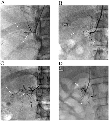
Table 3. Ratio of cortisol concentration of RAV and its primary branches to peripheral blood samples.
Table 4. Number of signature veins displayed during right adrenal venography with 4F MPA1 catheter (n = 53).
Data availability statement
The datasets generated or analyzed during the study are available from the corresponding author upon reasonable request.