
Figures & data
Figure 1. Group II patients without perioral duct tape fixation.
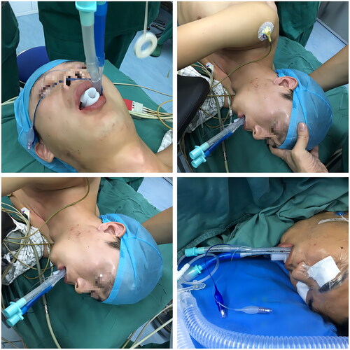
Figure 2. Group I patients with perioral duct tape fixation.
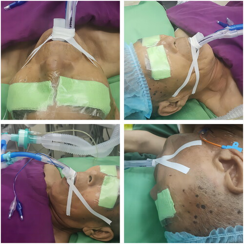
Figure 3. Study design drawing.
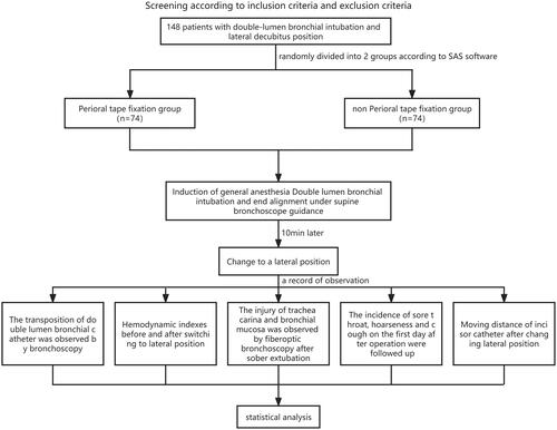
Table 1. The general characteristics of patients and comparison between the groups.
Figure 4. The cuff end dislocation and misalignment of the DLT after lateral Decubitus position in the two groups (n = 74).
Note: Compared with group I, αP < 0.05.
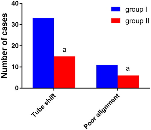
Figure 5. Hemodynamic changes before and after changing to a lateral position in both groups (± s), n = 74.
Note: Compared with T0, αP < 0.05.
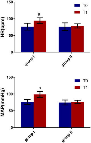
Figure 6. Comparison of injury to the tracheal carina and bronchial mucosa (n, %), n = 74.
Note: Compared with group, αP < 0.05.
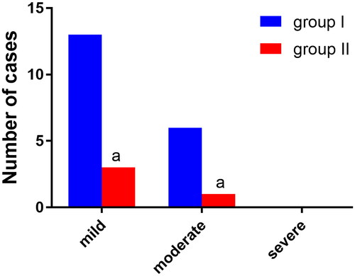
Figure 7. This is a Photograph of the tracheal mucosa of the patient in group I. it can be seen that the cuff end of the DLT is displaced outward, and the carinal mucosa is damaged, bleeding, and edema due to repeated catheter displacement.
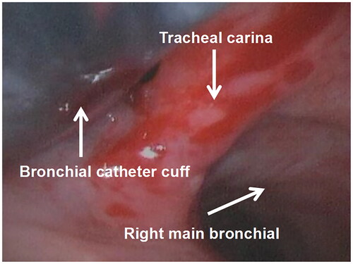
Figure 8. This is a Picture of the tracheal mucosa in group II, and it can be seen that the carinal and main bronchial mucosa have very little hemorrhage, damage, or edema.
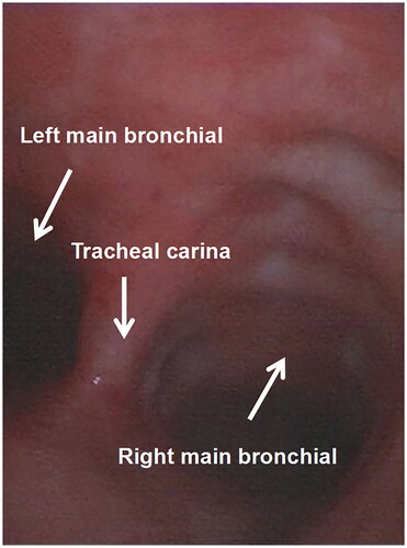
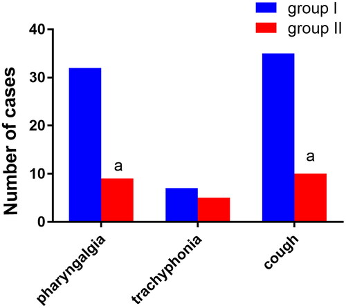
Figure 9. Comparison of postoperative sore throat, hoarseness, and cough between the two groups of patients (n, %), n = 74.
Note: Compared with group I, αP < 0.05.
Data availability statement
The datasets used and/or analysed during the current study available from the corresponding author on reasonable request.