
Figures & data
Figure 1. Hb analysis of simple heterozygosity of Hb Doi-Saket identified in the proband (A–C) and double heterozygosity of Hb Doi-Saket and HbE identified in his son (D–F). A and D show the Electropherograms; Hb Doi-Saket concealed HbF at electrophoretic zone 7. B and E are HPLC chromatograms analyzed using the VARIANT II system; Hb Doi-Saket was eluted by overlapping with HbA2 with a retention time of 3.92 min. C and F represent the HPLC chromatograms obtained using the Premier Resolution system; Hb Doi-Saket completely overlapped with HbA2 during elution.
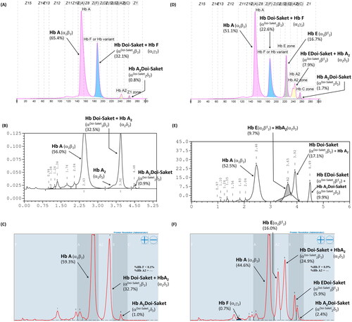
Table 1. Hematological data and globin genotypes of the proband and his family members.
Figure 2. Hb Separation profiles of the heterozygous Hb Q-Thailand using (A) capillary electrophoresis, (B) VARIANT II HPLC, and (C) Premier Resolution HPLC.
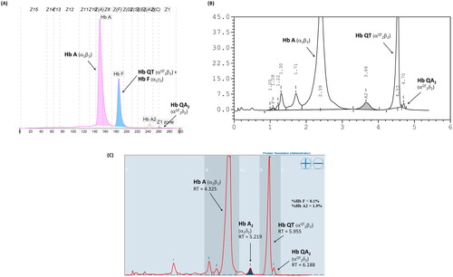
Figure 3. Locations of the four primers, the breakpoints of -α3.7 and -α4.2 deletions, and amplified DNA fragment. The 1,529- and 1,779-bp fragments are specific for -α3.7 and -α4.2, respectively. M, 100-bp marker; lane 1; normal control; lane 2, -α3.7 carrier; lane 3, -α4.2 carrier; lane 4, mother; lane 5, father; lane 6, proband; lane 7, wife of proband; and lane 8, son of proband.
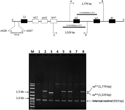
Figure 4. (A) the locus chromosome with -α3.7 deletion and forward directional nucleotide sequence obtained from the α2α1 hybrid and α1 genes. (B) the lack of deletion on the locus chromosome and forward directional nucleotide sequence obtained from the α2 gene. The arrow indicates replacement of the heterozygote nucleotide (codon 9 AAC>AAA), and any identical nucleotide substitution in the same place was not characterized in the α2 gene.
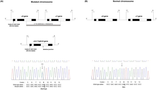
Figure 5. The hybrid -α3.7 gene and forward directional nucleotide sequence obtained from this gene. The arrow indicates nucleotide replacement.
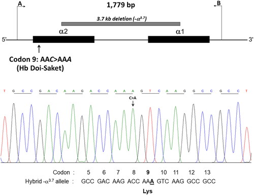
Figure 6. Identification of AAC>AAA mutation by HpyCH4IV digestion of the PCR product. (A) HpyCH4IV digestion of the in cis α2α1 hybrid and α1 genes. Lane 1 is the undigested amplified DNA (975 bp). lanes 2 and 5 are HpyCH4IV-digested amplified DNA of the proband’s mother and wife, who had normal α-globin allele (αα/αα). the 561-bp digested fragment in lanes 3, 4, and 6 indicates the presence of Hb Doi-Saket mutation in the proband, his father, and son. (B) HpyCH4IV digestion of the α2 gene. Three fragments of 506, 306, and 255 bp were produced, whereas the 561-bp fragment was not produced owing to the absence of AAC>AAA mutation.
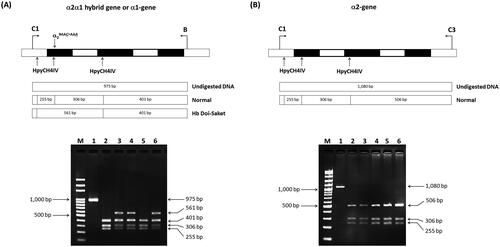
Figure 7. A multiplex ASPCR Assay for simultaneous identification of Hb Doi-Saket and Hb Q-Thailand. The locations and orientations of primers are indicated. The 729-bp fragment generated using SP42 and B is specific for αDoi-Saket, whereas the 416-bp fragment generated using αG20 and B is specific for αQ-Thailand. The 199-bp fragment is an internal control generated using SP44 and B. M, 100-bp ladder; lane 1, normal DNA; lane 2, DNA control of Hb Q-Thailand; lane 3, mother; lane 4, father; lane 5, proband; lane 6, wife of proband; lane 7, son of proband; and lane 8, Hb Q-Thailand carrier.
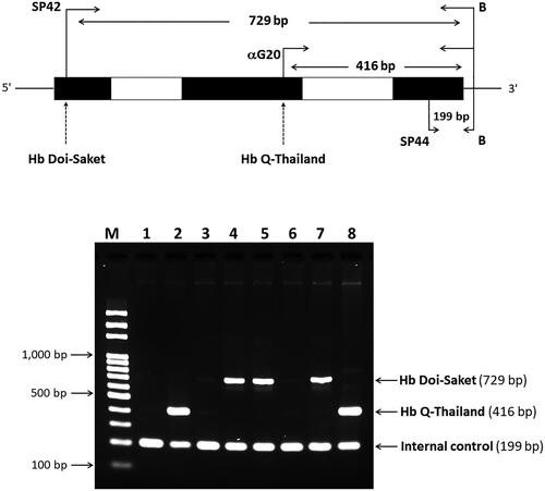
Table 2. α-Globin gene haplotypes associated with Hb Doi-Saket.
Data availability statement
The data that support the findings of this study are available from the corresponding author upon reasonable request.