
Figures & data
Table 1. Prevalence of skin disease in different military deployments.
Table 2. Prevalence of skin disease in non-deployed settings.
Figure 1. Cutaneous abscess on the lateral thigh. There is erythema, fluctuance, and purulence. Photo by Bruno Coignard, M.D.; Jeff Hageman, M.H.S. Reprinted from public domain Centers for disease Control and Prevention public Health Image Library; Retrieved from https://phil.cdc.gov/details.aspx?pid=7826.
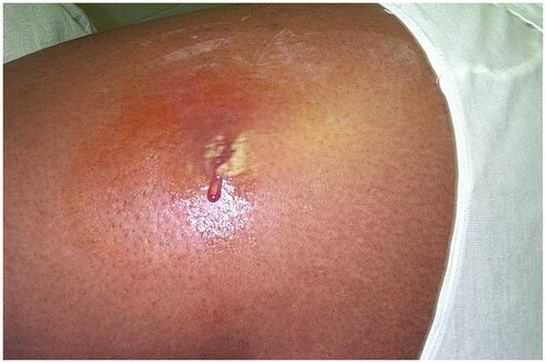
Figure 2. Tinea pedis. Annular scaly plaques involving the dorsal foot and web spaces. Reprinted with permission from: Oumeish and Parish [Citation40], with permission from Elsevier.
![Figure 2. Tinea pedis. Annular scaly plaques involving the dorsal foot and web spaces. Reprinted with permission from: Oumeish and Parish [Citation40], with permission from Elsevier.](/cms/asset/18c0bbdc-d5a7-429a-b014-e8f4d75ecb54/iann_a_2267425_f0002_c.jpg)
Figure 3. Cutaneous leishmaniasis. Central domed, eroded, and crusted erythematous nodules on the right dorsal hand of a soldier returning from Iraq deployment. This case has been previously reported. Reprinted with permission from Pehoushek et al. [Citation51], with permission from Elsevier.
![Figure 3. Cutaneous leishmaniasis. Central domed, eroded, and crusted erythematous nodules on the right dorsal hand of a soldier returning from Iraq deployment. This case has been previously reported. Reprinted with permission from Pehoushek et al. [Citation51], with permission from Elsevier.](/cms/asset/1d11b9e0-9324-4505-be08-ed627e6b38ec/iann_a_2267425_f0003_c.jpg)
Figure 4. Cutaneous leishmaniasis. Ulcerated plaque on the right fourth toe and third web space. This case has been previously reported. Reprinted with permission from: Pehoushek et al. [Citation51], with permission from Elsevier.
![Figure 4. Cutaneous leishmaniasis. Ulcerated plaque on the right fourth toe and third web space. This case has been previously reported. Reprinted with permission from: Pehoushek et al. [Citation51], with permission from Elsevier.](/cms/asset/9e73d3a1-796c-430c-800b-0132095ff87a/iann_a_2267425_f0004_c.jpg)
Figure 5. Scabies. Erythematous papules on the abdomen due to hypersensitivity to the scabies mite. Image by Joe Miller. Reprinted from public domain Centers for disease Control and Prevention public Health Image Library; Retrieved from https://phil.cdc.gov/details.aspx?pid=15382.
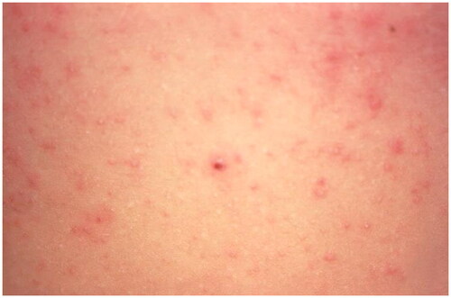
Figure 6. Cutaneous myiasis. Erythematous nodule with Central pustulation (a) and larva of C. anthropophaga, 7 mm long with an oval body and numerous black spines (b). Reprinted with permission from: Deng et al. [Citation74], with permission from John Wiley and Sons.
![Figure 6. Cutaneous myiasis. Erythematous nodule with Central pustulation (a) and larva of C. anthropophaga, 7 mm long with an oval body and numerous black spines (b). Reprinted with permission from: Deng et al. [Citation74], with permission from John Wiley and Sons.](/cms/asset/04c9b87e-7aca-4bab-96f2-a81f44c3bf88/iann_a_2267425_f0006_c.jpg)
Figure 7. Monkeypox. Umbilicated papules on the penile shaft. Reprinted with permission from: Vallée et al. [Citation96], with permission from Elsevier.
![Figure 7. Monkeypox. Umbilicated papules on the penile shaft. Reprinted with permission from: Vallée et al. [Citation96], with permission from Elsevier.](/cms/asset/db9e71f4-c544-49ce-bb34-cd5d9e63c84a/iann_a_2267425_f0007_c.jpg)
Figure 8. Pseudofolliculitis Barbae. Follicular papules and hyperpigmented patches. Reprinted from public domain United States Government document Tshudy and Cho [Citation131].
![Figure 8. Pseudofolliculitis Barbae. Follicular papules and hyperpigmented patches. Reprinted from public domain United States Government document Tshudy and Cho [Citation131].](/cms/asset/ed0b3bad-7384-4b78-ba41-1cf9fbcf86b6/iann_a_2267425_f0008_c.jpg)
Figure 9. Contact dermatitis of the leg and dorsal foot secondary to military boots. Erythematous plaques correspond to the outline of the military boots. Reprinted with permission from: Oumeish and Parish [Citation40], with permission from Elsevier.
![Figure 9. Contact dermatitis of the leg and dorsal foot secondary to military boots. Erythematous plaques correspond to the outline of the military boots. Reprinted with permission from: Oumeish and Parish [Citation40], with permission from Elsevier.](/cms/asset/d0d0214f-23b7-4c1e-a9f7-8e99a3494ad7/iann_a_2267425_f0009_c.jpg)
Figure 10. Frostbite of the lower leg and foot. Reprinted with permission from: Oumeish and Parish [Citation40], with permission from Elsevier.
![Figure 10. Frostbite of the lower leg and foot. Reprinted with permission from: Oumeish and Parish [Citation40], with permission from Elsevier.](/cms/asset/1ca9a10f-2fb6-4ee6-89f0-f03586b4723d/iann_a_2267425_f0010_c.jpg)
Data availability statement
Data sharing is not applicable to this article as no new data were created or analyzed in this study.