
Figures & data
Figure 1. Bladder tumor morphology under WLI and NBI modality. (A and B) Cauliflower-like lesions. These resembled a cauliflower and were primarily spherical or ellipsoidal, usually with a tip. (C and D) Papillary lesions. These were shorter cylindrical mucosal protrusions with a broad base that resembled a papilla. These may be present singly or as multiple short cylinders closely adjacent to each other in a cluster. For these lesions, there was a lack of a three-dimensional spherical structure when compared with cauliflower swellings. (E and F) Follicular lesions. Follicular lesions which were mainly light red or white vesicular structures under WLI, with thin and transparent walls and sparse vascularity visible within the vesicular structures. (G and H) Patchy lesions. Patchy lesions: slightly elevated or non-elevated lesion with an irregular distribution pattern and interspersed presence of lesion and normal mucosa. (I and J) Round lesions. Round lesions which were slightly elevated or non-elevated lesions with a broad base and a regular shape approximating a circle. (K and L) Villous lesions. Villous lesions featuring a thin cylindrical swelling that was slightly longer in length than the papillary lesions which were arranged in a column or a cluster and floated downstream under the influence of water, resembling a villus. (M and N) Flat bulges. Flat bulges which represented minor hemispherical swellings with a broad base, without a tip, and were elevated on the mucosal surface. These may be present singly or diffusely distributed in the bladder wall. (O and P) Mossy lesions. Mossy lesions which were slightly elevated lesions with a uniform thickness, the majority of which exhibit a lamellar or large lamellar distribution, resembling moss, with clear demarcation around the lesion and normal mucosa. These can be differentiated from patchy masses by the absence of regular mucosal interlacing within the area of the lesion. (Q and R) Algae-like lesions. Algae-like lesions which featured long thick stripes or long columnar swellings with tips. These can float downstream under the influence of water, resembling kelp, or with thick branches in a coral-like manner.
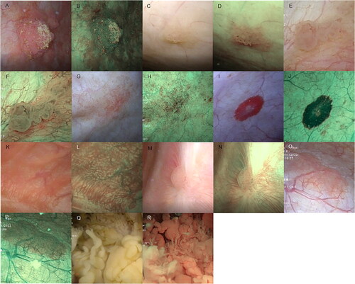
Figure 2. Illustration of the vascular morphology of the bladder mucosa by NBI modality. (A) Fine branching vessels. (B) Thick branching vessels. (C) Reticulated vessels. (D) Disorganized vessels. (E) Dotted vessels. (F) Circumferential vessels. (Red arrow indicates vessel location)
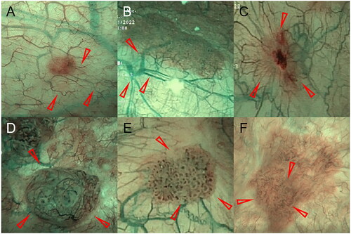
Figure 3. Delineation of the mucosal and basal mucosal areas of the lesions under NBI modality. (A) The yellow area represents the mucosal region of a cauliflower-like lesion with dotted vessels visible under the mucosa. Between the red and yellow areas lies the basal mucosal area, with a sparse distribution of thick and fine branching vessels and continuous vascular pathways. (B) The yellow area is the round lesion mucosal area, and between the red and yellow areas is the basal mucosal area, with sparse, thick visible branching vessels and continuous vascular alignment.
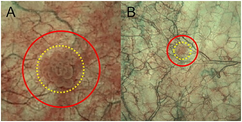
Table 1. Patient characteristics.
Supplemental Material
Download MS Word (100.5 KB)Data availability statement
The datasets used and/or analysed during the current study are available from the corresponding author on reasonable request.