
Figures & data
Table 1. Summary of common nail changes in older adults.
Figure 1. Pincer nail of the left first toenail in an 80-year-old woman. The lateral aspect of the nail plate is penetrating the periungual dermis of the lateral nail fold [Citation8].
![Figure 1. Pincer nail of the left first toenail in an 80-year-old woman. The lateral aspect of the nail plate is penetrating the periungual dermis of the lateral nail fold [Citation8].](/cms/asset/094f7e1e-611c-4d52-9fa6-46cdfe486c64/iann_a_2336989_f0001_c.jpg)
Figure 2. A 75-year-old female presented with painful bilateral great toenails for 10 years. Her nails grew slowly and were extremely difficult to clip. A full nail examination was significant for opaque yellow-brown thickening, hyperkeratosis, elongation, and increased curvature of the great toenails. Onychogryphosis can be differentiated from retronychia and onychomycosis by its spiral striated appearance [Citation13].
![Figure 2. A 75-year-old female presented with painful bilateral great toenails for 10 years. Her nails grew slowly and were extremely difficult to clip. A full nail examination was significant for opaque yellow-brown thickening, hyperkeratosis, elongation, and increased curvature of the great toenails. Onychogryphosis can be differentiated from retronychia and onychomycosis by its spiral striated appearance [Citation13].](/cms/asset/d6b599c8-c354-4d50-82e7-e036622296c5/iann_a_2336989_f0002_c.jpg)
Figure 3. Example of a patient-initiated nail hematoma selfie of the right thumbnail on the day of examination.
Patient-initiated nail hematoma selfie of the right thumbnail 1 month following the initial examination.
Patient-initiated nail hematoma selfie of the right thumbnail 2 months following the initial examination 18.
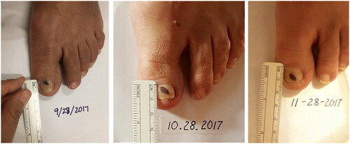
Figure 4. Clinical presentations of Beau’s lines, onychomadesis and retronychia. (A) Beau’s lines on the left toenails. (B) Onychomadesis of the left great toenail. (C) Retronychia of the right great toenail [Citation20].
![Figure 4. Clinical presentations of Beau’s lines, onychomadesis and retronychia. (A) Beau’s lines on the left toenails. (B) Onychomadesis of the left great toenail. (C) Retronychia of the right great toenail [Citation20].](/cms/asset/a81b51a8-59b9-4d92-8e02-3529b430db07/iann_a_2336989_f0004_c.jpg)
Figure 5. 93-year-old female with bullous pemphigoid presented with Beau’s lines on all fingernails at even intervals coinciding with her monthly IVIG treatments [Citation23].
![Figure 5. 93-year-old female with bullous pemphigoid presented with Beau’s lines on all fingernails at even intervals coinciding with her monthly IVIG treatments [Citation23].](/cms/asset/be83cd3f-3770-4dad-b683-e0480f77aec9/iann_a_2336989_f0005_c.jpg)
Figure 6. Physical examination findings in onychomycosis. A, Right great toenail with subungual hyperkeratosis and nail plate onycholysis. B, Left great toenail with yellow discoloration and onycholysis. C, Multiple toenails with subungual hyperkeratosis and onycholysis. D, Toenails with severe onychodystrophy and ridging. E, Scale on the plantar feet and web spaces [Citation33].
![Figure 6. Physical examination findings in onychomycosis. A, Right great toenail with subungual hyperkeratosis and nail plate onycholysis. B, Left great toenail with yellow discoloration and onycholysis. C, Multiple toenails with subungual hyperkeratosis and onycholysis. D, Toenails with severe onychodystrophy and ridging. E, Scale on the plantar feet and web spaces [Citation33].](/cms/asset/1607e130-713b-4045-8846-c80399380fe1/iann_a_2336989_f0006_c.jpg)
Figure 7. Dermoscopy of onychomycosis. A, Fringed proximal margin of the onycholysis. B, Blurred yellow-orange-brown nail discoloration in longitudinal striae (the fading mimics Aurora Borealis). C, Distribution of the discoloration in longitudinal striae or round areas. D, Ruin-like appearance of the subungual scales that are white-yellow-orange in color. Photographs courtesy of Dr Maria Bianca Piraccini [Citation34].
![Figure 7. Dermoscopy of onychomycosis. A, Fringed proximal margin of the onycholysis. B, Blurred yellow-orange-brown nail discoloration in longitudinal striae (the fading mimics Aurora Borealis). C, Distribution of the discoloration in longitudinal striae or round areas. D, Ruin-like appearance of the subungual scales that are white-yellow-orange in color. Photographs courtesy of Dr Maria Bianca Piraccini [Citation34].](/cms/asset/4d31402f-1d28-492d-af65-86a5f974be6a/iann_a_2336989_f0007_c.jpg)
Figure 8. Chronic paronychia presenting with edema of the right third and fourth nail folds. Of note, there is also benign longitudinal melanonychia of the right 4th fingernail.
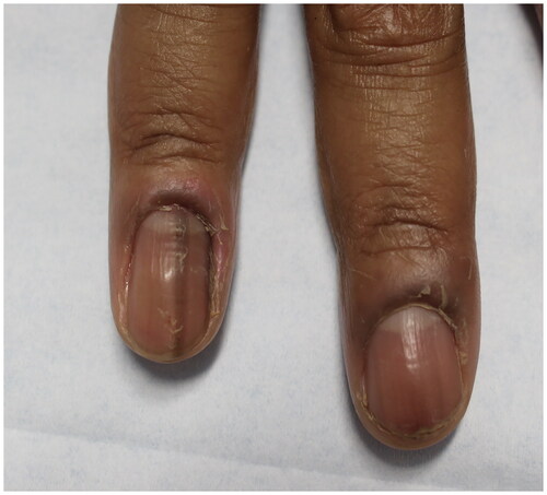
Figure 9. Clinical manifestations of brittle nail syndrome, including lamellar onychoschizia.
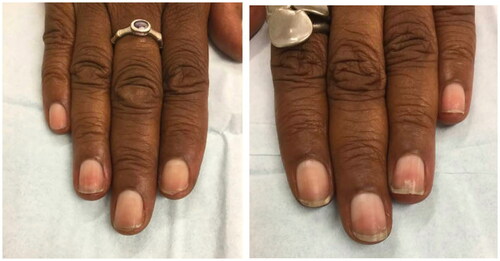
Figure 10. Nail pitting and onycholysis in right fingernails [Citation20].
![Figure 10. Nail pitting and onycholysis in right fingernails [Citation20].](/cms/asset/62b0e8bd-cbaf-424c-836a-f0c2ecf6f1a4/iann_a_2336989_f0010_c.jpg)
Figure 11. Dermoscopic appearance of subungual melanomas. B, Brown lines on a brown background, irregular color, thickness, and spacing with no loss of parallelism. C, Brown lines on a brown background, irregular color, thickness, and spacing with loss of parallelism[Citation62].
![Figure 11. Dermoscopic appearance of subungual melanomas. B, Brown lines on a brown background, irregular color, thickness, and spacing with no loss of parallelism. C, Brown lines on a brown background, irregular color, thickness, and spacing with loss of parallelism[Citation62].](/cms/asset/59a19bdd-c826-4219-9e33-3a9402f50375/iann_a_2336989_f0011_c.jpg)
Figure 12. A, A translucent compressible nodule of the proximal nail fold and longitudinal groove in the nail plate of the right thumb. B, Transillumination using a dermatoscope to project light from the dorsal digit through the nail unit demonstrated a central nodule in the proximal nail fold as well as a second cyst radially. (Reprinted with permission from Cutis. 2020;105(2):82. ©2020, Frontline Medical Communications Inc.) [Citation73].
![Figure 12. A, A translucent compressible nodule of the proximal nail fold and longitudinal groove in the nail plate of the right thumb. B, Transillumination using a dermatoscope to project light from the dorsal digit through the nail unit demonstrated a central nodule in the proximal nail fold as well as a second cyst radially. (Reprinted with permission from Cutis. 2020;105(2):82. ©2020, Frontline Medical Communications Inc.) [Citation73].](/cms/asset/62f3092f-2892-4883-be99-18ff579b8677/iann_a_2336989_f0012_c.jpg)
Data availability statement
Data sharing is not applicable to this article as no new data were created or analyzed in this study