Figures & data
Table I. Demographic and clinical characteristics of the study population (n = 107).
Table II. Biochemical characteristics and other study variables.
Figure 1. Box plots showing median(horizontal lines), 25th to 75th percentiles (boxes), and 95th percentiles (whiskers) in the different groups by global percent diameter stenosis index (A), global extent index (B), and global atheroma burden index (C), respectively. P12 value indicates comparison between group 1 (HOMA IR <1.8) and group 2 (HOMA IR ⩾1.8) and P13 value between group 1 (HOMA IR <1.8) and group 3 (diabetic subjects). P‐values from analysis of covariance with adjustment for age and gender. For definition of indexes, see Methods.
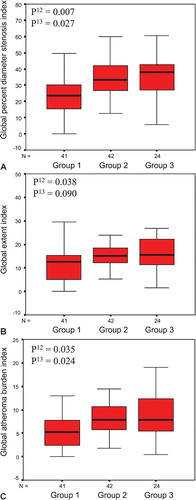
Table III. Quantitative coronary angiography results.
Table IV. Multivariate regression model of global percent diameter stenosis index.