Figures & data
Figure 1 Endothelial progenitor cells and endothelial colony forming units under light microscopy. Note that the size of colony forming units(CFUs) varies widely. a: small CFUs consisting of a few cells (40×); b and c: bigger CFUs (40×, 100×); d: predominantly single located endothelial cells without accumulation in CFUs (40×).
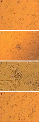
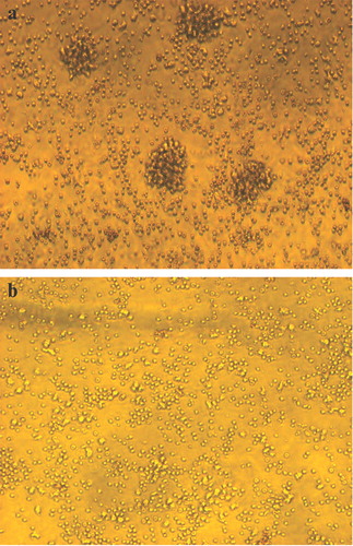
Figure 2 Endothelial progenitor cells(EPCs) migration and colony forming units (CFUs). a: Migration of EPCs to each other before forming CFUs (40×). b: This process may be impaired in some patients (40×).