681
Views
40
CrossRef citations to date
0
Altmetric
Review Article

Management of inherited von Willebrand disease in 2007

&
Pages 346-358 | Published online: 08 Jul 2009

Figures & data

Figure 1. Schematic representation of the von Willebrand factor(VWF) gene located in chromosome 12: the main exons are indicated with the number of base pairs from 5′ to 3′ (upper panel). The structure of VWF functional domains: the pre‐pro‐VWF is indicated with amino acids numbered from the amino‐ (aa 1) to carboxy‐terminal portions (aa 2813) of VWF. Note the important CK and D3 domains for formation of VWF dimers and multimers. The native mature subunit of VWF, after the cleaving of the pre‐pro‐VWF, is described with its functional domains: the VWF binding sites for factor VIII (D′ and D3), GPIb, botrocetin, heparin, sulfatide, collagen (A1), collagen (A3) and the arginine, glycine, aspartic acid (RGD) sequence for binding to αIIbβ3 (middle panel). Distribution of VWF mutations in patients with VWD types 2: the positions of mutations causing VWD types 2A, 2B, 2M, 2N are indicated with black bars throughout the VWF domains (lower panel).
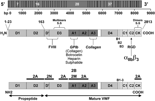
Figure 1. Schematic representation of the von Willebrand factor(VWF) gene located in chromosome 12: the main exons are indicated with the number of base pairs from 5′ to 3′ (upper panel). The structure of VWF functional domains: the pre‐pro‐VWF is indicated with amino acids numbered from the amino‐ (aa 1) to carboxy‐terminal portions (aa 2813) of VWF. Note the important CK and D3 domains for formation of VWF dimers and multimers. The native mature subunit of VWF, after the cleaving of the pre‐pro‐VWF, is described with its functional domains: the VWF binding sites for factor VIII (D′ and D3), GPIb, botrocetin, heparin, sulfatide, collagen (A1), collagen (A3) and the arginine, glycine, aspartic acid (RGD) sequence for binding to αIIbβ3 (middle panel). Distribution of VWF mutations in patients with VWD types 2: the positions of mutations causing VWD types 2A, 2B, 2M, 2N are indicated with black bars throughout the VWF domains (lower panel).

Table I. Prevalence (%) of bleeding symptoms in patients with VWD from different cohorts and in normal individuals (adapted from Federici Citation12, Silwer Citation17, Lak et al. Citation18).

Table II. Bleeding score used to evaluate the bleeding history (see Tosetto et al. Citation20).

Table III. Clinical and laboratory parameters used for VWD diagnosis.

Figure 2. Flowchart proposed for the diagnosis of different von Willebrand disease(VWD) types. Type 3 VWD can be diagnosed in case of unmeasurable VWF:Ag (a). A proportionate reduction of both VWF:Ag and VWF:RCo with a RCo/Ag ratio >0.7 suggests type 1 VWD (b). If the VWF:RCo/Ag ratio is <0.7 type 2 is diagnosed. Type 2B VWD (d) can be identified in case of heightened ristocetin‐induced platelet aggregation (RIPA) (<0.8 mg/mL), whereas types 2A and 2M cause low RIPA (>1.2 mg/mL). Multimeric analysis in plasma (e) is necessary to distinguish between type 2A VWD (lack of the largest and intermediate multimers) and type 2M VWD (all the multimers present). Type 2N VWD can be suspected in case of discrepant values for FVIII (c) and VWF:Ag (ratio <0.5), and diagnosis should be confirmed by the specific test (g) of VWF:factor VIII binding capacity (VWF:FVIIIB). In type 1 VWD the ratio between Factor VIII and VWF:Ag is always ⩾1 and the severity of the type 1 VWD phenotype can usually be evaluated from platelet VWF (f) measurements. (This figure was derived from that originally reported previously Citation12.)
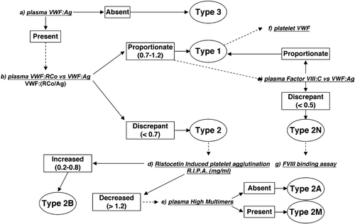
Figure 2. Flowchart proposed for the diagnosis of different von Willebrand disease(VWD) types. Type 3 VWD can be diagnosed in case of unmeasurable VWF:Ag (a). A proportionate reduction of both VWF:Ag and VWF:RCo with a RCo/Ag ratio >0.7 suggests type 1 VWD (b). If the VWF:RCo/Ag ratio is <0.7 type 2 is diagnosed. Type 2B VWD (d) can be identified in case of heightened ristocetin‐induced platelet aggregation (RIPA) (<0.8 mg/mL), whereas types 2A and 2M cause low RIPA (>1.2 mg/mL). Multimeric analysis in plasma (e) is necessary to distinguish between type 2A VWD (lack of the largest and intermediate multimers) and type 2M VWD (all the multimers present). Type 2N VWD can be suspected in case of discrepant values for FVIII (c) and VWF:Ag (ratio <0.5), and diagnosis should be confirmed by the specific test (g) of VWF:factor VIII binding capacity (VWF:FVIIIB). In type 1 VWD the ratio between Factor VIII and VWF:Ag is always ⩾1 and the severity of the type 1 VWD phenotype can usually be evaluated from platelet VWF (f) measurements. (This figure was derived from that originally reported previously Citation12.)

Table IV. List of the most frequent mutations in type 2A, 2B, 2M, and 2N according to VWF domains and exons of VWF gene.

Table V. Recommendations for the infusion test with DDAVP

Table VI. Treatment of different types of VWD.

Reprints and Corporate Permissions

Please note: Selecting permissions does not provide access to the full text of the article, please see our help page How do I view content?

To request a reprint or corporate permissions for this article, please click on the relevant link below:

Academic Permissions

Please note: Selecting permissions does not provide access to the full text of the article, please see our help page How do I view content?

Obtain permissions instantly via Rightslink by clicking on the button below:

If you are unable to obtain permissions via Rightslink, please complete and submit this Permissions form. For more information, please visit our Permissions help page.