Figures & data
Table I. Characteristics of the 12 Russian‐Karelian children who tested positive for tTGA in the primary screening; 8 of these children underwent endoscopy and small‐bowel biopsy to confirm the diagnosis of celiac disease (cases 1–8), while 4 children did not undergo endoscopy (cases 9–12).
Figure 1 Screening protocol for celiac disease based on the measurement of IgA‐class tissue transglutaminase antibodies (tTGA) in the two study populations followed by small‐bowel biopsy in tTGA‐positive subjects.
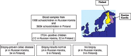
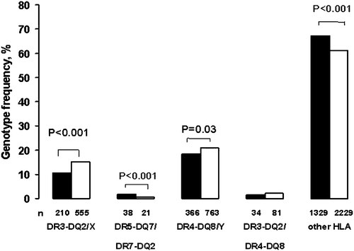
Figure 2 Frequency of different human leukocyte antigen(HLA) genotypes in schoolchildren in Russian Karelia (black bars) and in Finland (white bars). Altogether 1977 Russian‐Karelian and 3649 Finnish children were HLA‐typed (X = other than DQ7 or DQ8; Y = other than DQ2; n = absolute number of children carrying different HLA genotypes).