Figures & data
Table I. Acute and subacute complications after pacemaker or implantable cardioverter-defibrillator implantation in 150 patients.
Figure 1. Venographic images prior to implantation of a dual chamber pacemaker (A), and 6 months (B) and 25 months (C) postoperatively on a 57-year-old male patient. A stenosis (arrow) of the subclavian vein was seen in the 6-month study (B), and complete occlusion of the same vessel with collateral venous flow after 2 years (C).
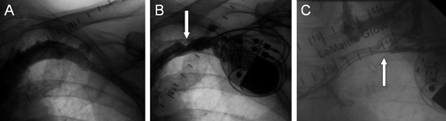
Figure 2. Panels A and B depict a filling defect suggestive of thrombus formation (arrows) in the 6-month venography of an 81-year-old male patient implanted with a biventricular pacemaker. Late venography (C) 29 months after implantation shows complete occlusion of the axillary vein (arrow) and several new collateral vessels.
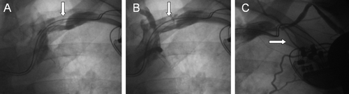
Table II. Venographic findings in 50 patients at base-line, 6 months, and at 2 years.