1,443
Views
4
CrossRef citations to date
0
Altmetric
Original Articles

Embryonic and larval development of the peanut worm Phascolosoma agassizii (Keferstein 1867) from the Sea of Japan (Sipuncula: Phascolosomatidea)

, &
Pages 22-29 | Received 03 Feb 2010, Accepted 01 Sep 2010, Published online: 02 Feb 2011

Figures & data

Figure 1. Embryonic stages and larvae of P. agassizii. A: 20 min; B: 40 min; C: 1 h 40 min; D: 2 h 10 min; E: 2 h 40 min; F: 3 h 10 min; G: 4 h 10 min; H: 6 h 30 min; I: 8 h; J: 24 h, lateral view; K: 48 h, frontal view; L: 48 h, lateral view. Numerals indicate serial numbers of blastomeres. AO, apical organ; AR, archenteron; BP, blastopore; DD, descendant of 2d blastomere; EB, entodermal blastomeres; EN, entoderm; MB, mesoblast; MD, mesoderm; MO, mouth; PH, pharynx; PR, prototroch; RA, rudiment of apical organ.
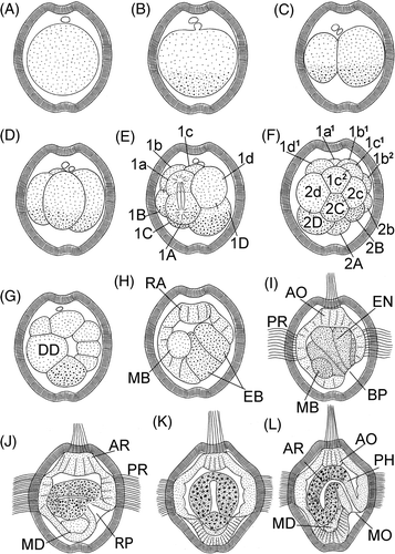
Figure 1. Embryonic stages and larvae of P. agassizii. A: 20 min; B: 40 min; C: 1 h 40 min; D: 2 h 10 min; E: 2 h 40 min; F: 3 h 10 min; G: 4 h 10 min; H: 6 h 30 min; I: 8 h; J: 24 h, lateral view; K: 48 h, frontal view; L: 48 h, lateral view. Numerals indicate serial numbers of blastomeres. AO, apical organ; AR, archenteron; BP, blastopore; DD, descendant of 2d blastomere; EB, entodermal blastomeres; EN, entoderm; MB, mesoblast; MD, mesoderm; MO, mouth; PH, pharynx; PR, prototroch; RA, rudiment of apical organ.

Figure 2. Larvae of P. agassizii. A: 68 h, frontal view; B: 80 h, frontal view; C: 80 h, lateral view. Arrowheads indicate mouth opening. AO, apical organ; CM, coelomic mesoderm; CO, coelom; EY, eyespot; GU, thin gut; PH, pharynx; PR, prototroch; RB, rudiment of buccal organ; RH, rudiment of hindgut; ST, stomach.
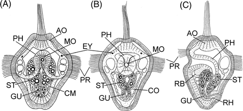
Figure 2. Larvae of P. agassizii. A: 68 h, frontal view; B: 80 h, frontal view; C: 80 h, lateral view. Arrowheads indicate mouth opening. AO, apical organ; CM, coelomic mesoderm; CO, coelom; EY, eyespot; GU, thin gut; PH, pharynx; PR, prototroch; RB, rudiment of buccal organ; RH, rudiment of hindgut; ST, stomach.

Figure 3. Embryonic stages and larvae of P. agassizii, light micrographs. A: 40 min; B: 1 h 20 min; C: 2 h 20 min; D: 3 h; E: 5 h; F: 7 h; G: 9 h; H: 12 h; I: 50 h; J: 60 h; K: 1 month, lateral view. Bars: 20 µ. AN, anus; AP, tuft of apical cilia; EY, eyespot; GU, thin gut; LI, lip; MO, mouth; MT, metatroch; PR, prototroch; TE, retracted terminal organ.
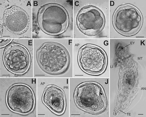
Figure 3. Embryonic stages and larvae of P. agassizii, light micrographs. A: 40 min; B: 1 h 20 min; C: 2 h 20 min; D: 3 h; E: 5 h; F: 7 h; G: 9 h; H: 12 h; I: 50 h; J: 60 h; K: 1 month, lateral view. Bars: 20 µ. AN, anus; AP, tuft of apical cilia; EY, eyespot; GU, thin gut; LI, lip; MO, mouth; MT, metatroch; PR, prototroch; TE, retracted terminal organ.

Figure 4. Embryonic stages and larvae of P. agassizii. SEM micrographs. A–G – stages rared in the laboratory, H – larvae from plankton. A–B, eggs; C–E, trochophores; F–H, pelagospheras. A: 2 h, lateral view; B: 2 h, view from vegetal pole; C: 9 h; D: 70 H; E: 80 h; F: 10 days; G: 15 days; H: 1 month. Bars: A–C 10 µ, D–G 20 µ, H 60 µ. White arrowhead indicates sensory groove. AD, apical depression; AN, anus; AP, tuft of apical cilia; DL, dorso-lateral ciliary lobes; LI, lip; MO, mouth; MT, metatroch; PR, prototroch; TP, trunk papillae; VD, vegetal depression.
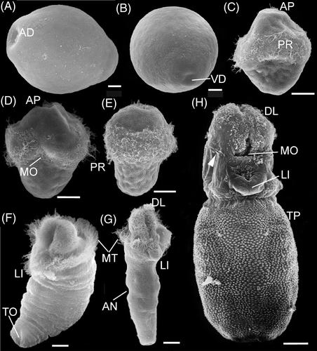
Figure 4. Embryonic stages and larvae of P. agassizii. SEM micrographs. A–G – stages rared in the laboratory, H – larvae from plankton. A–B, eggs; C–E, trochophores; F–H, pelagospheras. A: 2 h, lateral view; B: 2 h, view from vegetal pole; C: 9 h; D: 70 H; E: 80 h; F: 10 days; G: 15 days; H: 1 month. Bars: A–C 10 µ, D–G 20 µ, H 60 µ. White arrowhead indicates sensory groove. AD, apical depression; AN, anus; AP, tuft of apical cilia; DL, dorso-lateral ciliary lobes; LI, lip; MO, mouth; MT, metatroch; PR, prototroch; TP, trunk papillae; VD, vegetal depression.

Figure 5. Pelagospheras of P. agassizii. A: 96 h, lateral view; B: 96 h, frontal view; C: 120 h, lateral view; D: 180 h, frontal view; E: 180 h, lateral view; F: 1 month, lateral view. AO, apical organ; AN, anus; AS, apical sensilla; BO, buccal organ; BR, brain; CC, circular coelomic canal; CE, coelomocyte; CL, coelomic canals in dorsa-lateral lobes; DL, dorso-lateral ciliary lobes; DR, dorsal retractor; EY, eyespot; GU, thin gut; HG, hindgut; LG, lip gland; LI, lip; ME, mesenterium; MO, mouth; MT, metatroch; NE, nephridium; PH, pharynx; PO, sensory-glandular pore; PR, prototroch; PC, pharyngeal connectives; RN, ventral nerve cord; RP, rudiment of prototroch; RT, retractor of terminal organ; SP, sensory papilla; ST, stomach; TO, terminal organ; VR, ventral retractor.
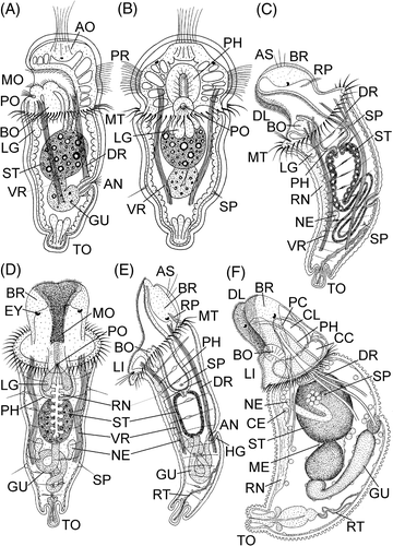
Figure 5. Pelagospheras of P. agassizii. A: 96 h, lateral view; B: 96 h, frontal view; C: 120 h, lateral view; D: 180 h, frontal view; E: 180 h, lateral view; F: 1 month, lateral view. AO, apical organ; AN, anus; AS, apical sensilla; BO, buccal organ; BR, brain; CC, circular coelomic canal; CE, coelomocyte; CL, coelomic canals in dorsa-lateral lobes; DL, dorso-lateral ciliary lobes; DR, dorsal retractor; EY, eyespot; GU, thin gut; HG, hindgut; LG, lip gland; LI, lip; ME, mesenterium; MO, mouth; MT, metatroch; NE, nephridium; PH, pharynx; PO, sensory-glandular pore; PR, prototroch; PC, pharyngeal connectives; RN, ventral nerve cord; RP, rudiment of prototroch; RT, retractor of terminal organ; SP, sensory papilla; ST, stomach; TO, terminal organ; VR, ventral retractor.

Reprints and Corporate Permissions

Please note: Selecting permissions does not provide access to the full text of the article, please see our help page How do I view content?

To request a reprint or corporate permissions for this article, please click on the relevant link below:

Academic Permissions

Please note: Selecting permissions does not provide access to the full text of the article, please see our help page How do I view content?

Obtain permissions instantly via Rightslink by clicking on the button below:

If you are unable to obtain permissions via Rightslink, please complete and submit this Permissions form. For more information, please visit our Permissions help page.