
Figures & data
Figure 1. Representative figure of deep white matter lesion (A) and periventricular white matter lesion (B) on a fluid-attenuated inversion recovery (FLAIR) magnetic resonance imaging.
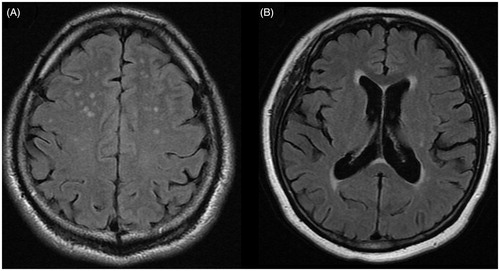
Table 1. Clinical, laboratory and treadmill exercise data for subjects with and without WMLs.
Table 2. Logistic regression analysis of potential risk factors for WMLs.
Table 3. Clinical, laboratory and treadmill exercise data for normotensive subjects with and without WMLs.
Table 4. Logistic regression analysis of potential risk factors for WMLs in normotensive individuals.