
Figures & data
Table 1. Orthokeratology care solutions from Ophtecs Corporation (Tokyo, Japan)
Table 2. Clinical findings from September 2018 to September 2019
Figure 1. Corneal topography indicating lens decentration. A: 15-month visit, B: 18-month visit. R-right eye, L-left eye. The Aladdin HW3.0 (Topcon Medical Systems. Inc., Oakland US) was used at 15-month visit, and the Medmont E300 (Version 6.1.2, Medmont Pty. Ltd., Camberwell Australia) was used at 18-month visit
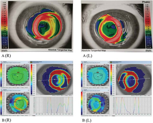
Figure 2. Ocular findings showing corneal A: microcystic responses, B: corneal haze, and C: corneal staining when first noted. R-right eye, L-left eye. Images captures using Topcon slit-lamp biomicroscopy (SL-D7, Topcon Medical Systems Inc., Oakland, USA)
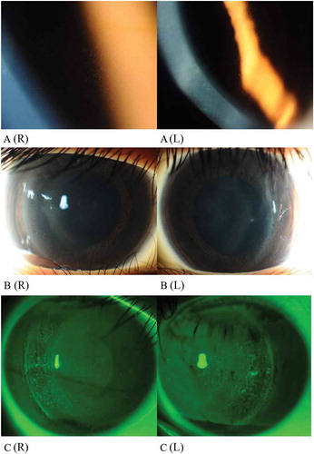
Table 3. Grading scale for orthokeratology lens decentration