Figures & data
Figure 1. Schematic of normal ageing changes, including biomechanical, vascular and neuronal changes. Understanding how such changes increase the risk of retinal ganglion cell injury may point to novel treatment approaches. RGC, retinal ganglion cell; RNFL, retinal nerve fibre layer; LC, lamina cribrosa.
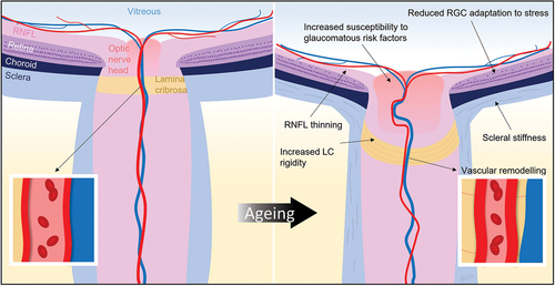
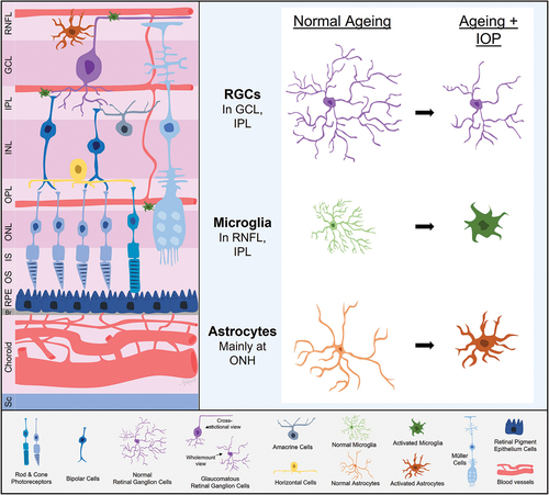
Figure 2. Retinal ganglion cell (RGC) and glial adaptations with ageing and intraocular pressure (IOP). With age, RGCs show reduced complexity along with evidence of an attenuated capacity for IOP induced morphological changes. Microglia, astrocytes and perhaps Müller cells in older eyes take on a more proinflammatory state and appear to show a slower but more exaggerated response to IOP elevation. RNFL, retinal nerve fibre layer; GCL, ganglion cell layer; IPL, inner plexiform layer; INL, inner nuclear layer; OPL, outer plexiform layer; ONL, outer nuclear layer; IS, photoreceptor inner segment layer; OS, photoreceptor outer segment layer; RPE, retinal pigment epithelium; Br, Bruch’s membrane; Sc, sclera; ONH, optic nerve head.