Figures & data
Figure 1. Anatomical representation of the peripheral fundus. The central zone is indicated by the dark pink inner circle (~30° diameter) and the equator is indicated by the blue circle (~50° diameter) which intersects the vortex veins.
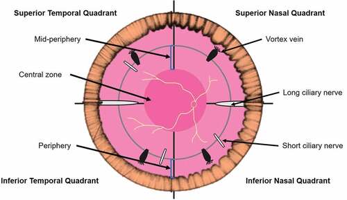
Figure 2. Dentate processes (arrow) visible in the temporal peripheral retina of a 31 year old Caucasian female using (A) UWF imaging, and separated as the (B) red-free image and (C) green-free image. A meridional fold is also present (asterisk). Abbreviations: UWF, ultra-widefield; RF, red-free; GF, green-free.
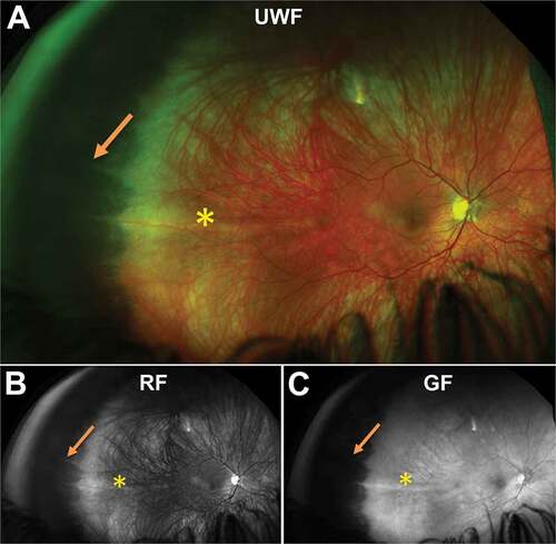
Figure 3. 31 year old Caucasian male with an oral bay and oral pearl in the far temporal peripheral retina visible on (A) UWF imaging and associated (B) red-free and (C) green-free images. (D) En face, infra-red imaging and (E) peripheral OCT through the structure demonstrates separation of the retinal layers at the bay. Abbreviations: IF, infra-red; OCT, optical coherence tomography. All other abbreviations as in .
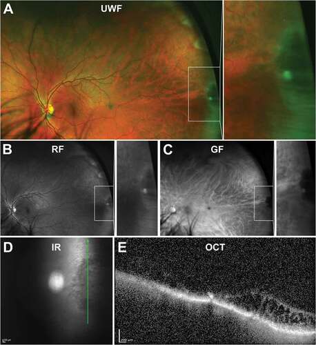
Figure 4. 66 year old Caucasian male meridional folds in the far temporal peripheral retina. White arrows highlight the folds within the magnified image. Abbreviations as in .
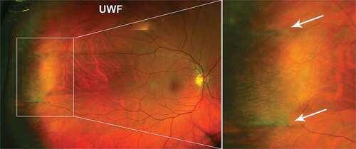
Figure 5. 32 year old Caucasian female with microcystoid degeneration. (A) UWF imaging demonstrates microcystoid degeneration in the infero - temporal peripheral retina and (B) red –free and (C) green-free images highlight the increased choroidal reflex due to the cysts. (D) On infra-red imaging, microcystoid degeneration can appear as a hazy area. (E) Peripheral OCT through the structure highlights separation of the inner retinal layers typical in reticular microcystoid degeneration. Abbreviations as in .
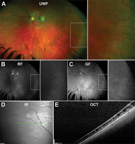
Figure 6. 87 year old Caucasian female with pavingstone degeneration. (A) UWF imaging demonstrates extensive lesions in the nasal retina which appear as (B) hypofluorescent lesions on FAF. (C) Red and (D) green-free images highlight the increased visibility of the choroid underneath lesions. (E) On infra-red imaging, pavingstone degeneration appears as hyporeflective lesions. (F) Peripheral OCT highlights thinning of the retina at lesions and subsequent increased choroidal visibility. Abbreviations as in .
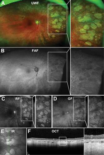
Figure 7. (A) UWF imaging of peripheral drusen in the nasal and temporal retina in a 66 year old Caucasian female which appear as (B) hyperfluorescent spots on FAF and (C) hyperreflective lesions on red-free and (D) green free imaging. (E) Infra-red imaging of peripheral drusen in a 56 year old Caucasian male which appear as reflective lesions. (F) OCT through peripheral drusen indicate similar morphology to macular drusen and location in the sub-RPE space. Abbreviations as in .
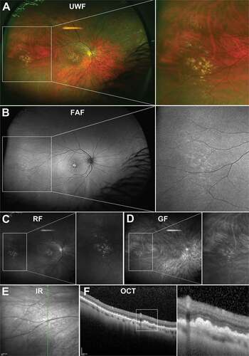
Figure 8. 73 year old Caucasian female with reticular pigmentary degeneration. (A) UWF imaging demonstrates reticular pigmentary degeneration in the nasal far periphery and magnification of this area demonstrates the typical “honeycomb” appearance. (B) On FAF, these structures can appear predominantly hypofluorescent. (C) Red-free and (D) green-free images highlight reticular pigmentary degeneration as light and dark areas respectively pointing to RPE involvement. (E) Infra-red imaging shows hyperreflective lesions corresponding to reticular pigmentary degeneration (F) OCT imaging highlights RPE involvement and outer retinal disruption of reticular pigmentary degeneration. Abbreviations as in .
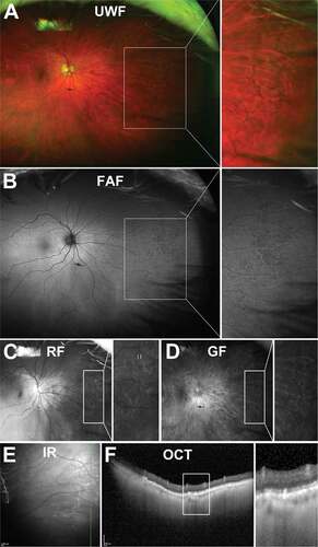
Figure 9. 22 year old Asian female with WWOP. (A) UWF imaging demonstrates irregular patches in the retinal periphery which are (B) hypofluorescent on FAF. Patches are visible on (C) red-free but less so on (D) green-free imaging. (E) Infrared imaging and (F) accompanying peripheral OCT at the margin of the lesion highlight the alterations to the ellipsoid zone in the outer retina. Abbreviations as in .
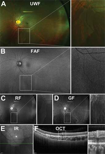
Figure 10. 29 year old Asian male with DWOP. (A) UWF imaging demonstrates irregular patches in the retinal periphery which show no alteration on (B) FAF but are visible on (C) red-free, less so on (D) green-free imaging. (E) Infrared imaging demonstrates DWOP as hyporeflective region and (F) peripheral OCT at the margin of the lesion highlights the alterations in reflectivity of the ellipsoid zone in the outer retina in DWOP. Abbreviations as in .
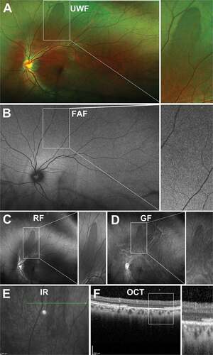
Figure 11. A 60-year-old East Asian male with lattice degeneration in the superior-temporal peripheral retina of the right eye. (A) UWF imaging highlights an oval-shaped area of whitened retina with irregular borders and internal RPE hyperplasia and (B) red-free and (C) green-free imaging suggests that the internal hyperpigmentation is located in the RPE layer with no choroidal involvement. (D) Infrared imaging demonstrates irregular reflectivity in the region of degeneration and shows the orientation of the lesion to be largely parallel to the ora serrata and (E) peripheral OCT, with the line scan orientated radially through the lesion, highlights the overlying vitreoretinal attachment at the anterior and posterior aspects and shows a retinal break at the anterior edge of the lesion, internal disorganisation of the retinal layers, and irregular hypo-reflectivity in the outer retinal layers likely related to the overlying vitreoretinal traction. Abbreviations as in .
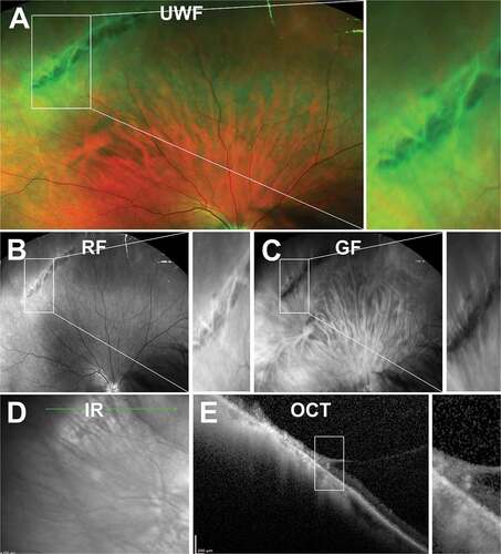
Figure 12. 29 year old Caucasian male with snail track degeneration in the superotemperal peripheral retina. (A) UWF imaging (B) red-free and (C) green-free imaging highlights closely spaced circular lesions forming a ‘track’. (D) Infra red imaging and associated (E) peripheral OCT highlights thinning and a wrinkled inner retina. In some cases, (F - G) traction can be observed. Abbreviations as in .
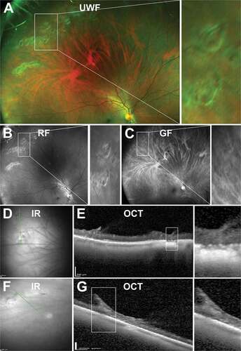
Figure 13. 86 year old Caucasian male with degenerative retinoschisis. (A) UWF, (B) red-free, (C) green-free and (D) infra-red imaging highlight the transparent dome-shaped elevation of the retina associated with retinoschisis. (E) Peripheral OCT highlights the splitting of the inner retinal layers and remnants of intraretinal pillars. Abbreviations as in .
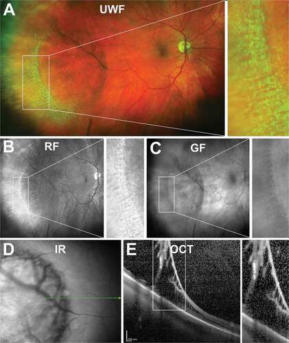
Figure 14. 84-year-old Caucasian male with operculated retinal hole. (A) UWF imaging, (B) red and (C) green-free and (D) infra-red imaging show a round lesion with a surrounding cuff and an adjacent operculum. (E) Peripheral OCT highlights the flat profile of the full-thickness hole with no associated subretinal fluid. Abbreviations as in .
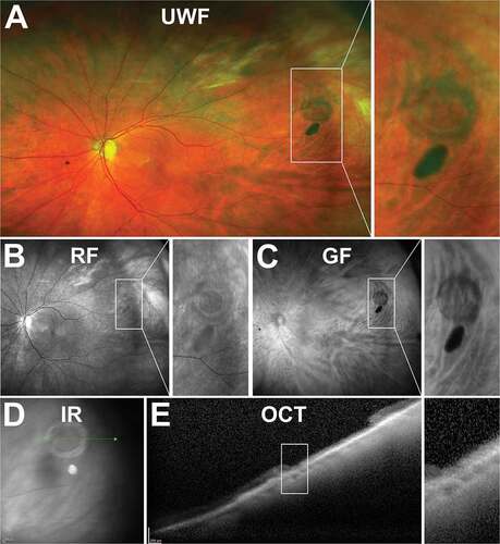
Figure 15. 23 year old Caucasian female with atrophic retinal hole within an area of lattice degeneration. (A) UWF imaging, (B) red and (C) green-free and (D) infra-red imaging show similar characteristics to operculated holes of a round lesion with a surrounding cuff. (E) Peripheral OCT highlights the full-thickness loss of retinal tissue at the site of the hole but no sign of overlying or displaced tissue. Abbreviations as in .
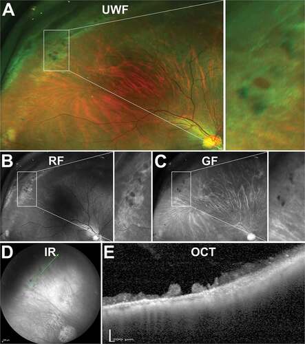
Figure 16. 47 year old Caucasian male with non-cystic vitreoretinal tuft. (A) UWF imaging indicates the well demarcated lesion in the superonasal peripheral retina, visible on (B) red-free and (C) green-free images. (D) On infrared imaging, the lesion appears dark and corresponding (E) peripheral OCT highlights the hyperreflective apical tip of the tuft extending into the vitreous with disturbance to the underlying photoreceptor and RPE layers. Abbreviations as in .
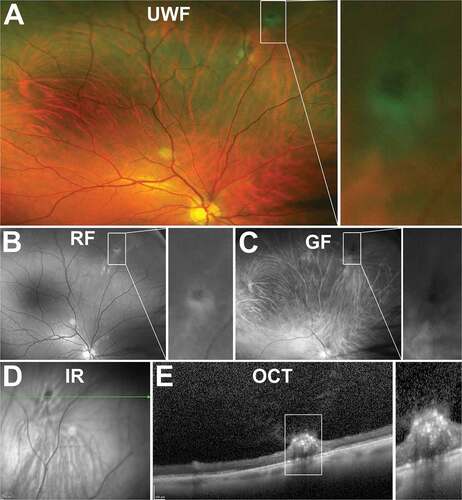
Figure 17. 43 year old Asian female with multiple cystic vitreoretinal tufts in the superior peripheral retina. (A) On UWF imaging, tufts appeared as bright, sharp lesions which remained visible on (B) red-free and (C) green-free images. (D) Infrared imaging shows lesions are hyporeflective and (E) peripheral OCT demonstrates tufts have a large, hyper-reflective apical tip extending into the vitreous with internal cystic spaces and loss of reflectivity of the underlying inner and outer retinal layers. Abbreviations as in .
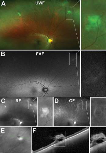
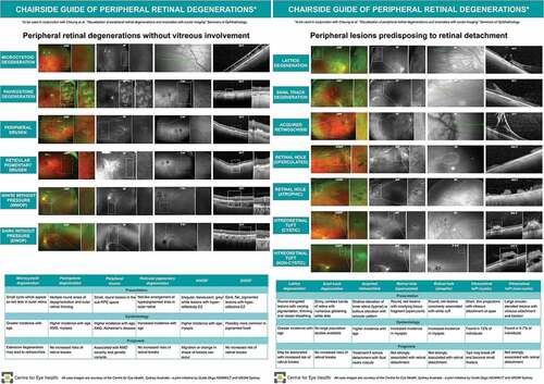
Figure 18. Chairside guide of peripheral retinal degenerations including summary of presentation, epidemiology (if known), prognosis and appearance with ocular imaging.