Figures & data
Figure 1. (a-f): Corneal topography of the contralateral eye of the patients; a- Patient 1, b-Patient 2, c-Patient 3, d-Patient 4, e-Patient-6, f-Patient 7 (Patient 5 with Down’s syndrome could not cooperate for topography).
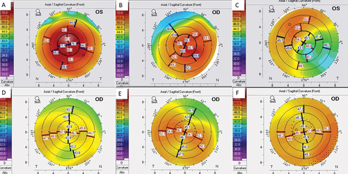
Figure 2. (a-c)- a: Slit-lamp photograph of the right eye when patient presented with a three-day history of acute hydrops; b: Slit-lamp photograph of the right eye after one month of conservative management showed marked increase in corneal edema; c: Slit-lamp photograph at first postoperative day showing rapid clearing of the peripheral cornea; d: Clinical picture on the fifth postoperative day showing further clearing of peripheral cornea. The optical coherence tomography pictures at first visit, one month after conservative treatment, postoperative day 1 and day 5 are depicted in e, f, g, and h, respectively.
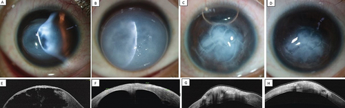
Figure 3. (a-c)- a: Slit-lamp photograph of the left eye when patient showing hydrops; b: Slit-lamp photograph of the left eye 1 h after the surgery shows significant resolution of peripheral corneal edema; c: Slit-lamp photograph at day 1 postoperative period.
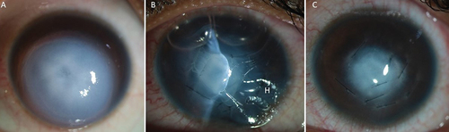
Figure 4. (a-f)- a: Slit-lamp photograph of the right eye when patient presented with a two-week history of increasing hydrops following blunt injury. The mid peripheral cornea shows circular area of differential transparency corresponding to posterior membrane dehiscence; b: Slit-lamp photograph of the right eye on first postoperative day shows remarkable resolution of corneal edema; c: Slit-lamp photograph at 1-week postoperative period shows improvement in corneal clarity; d-f: The corresponding optical coherence tomography pictures at pre-op, day 1 and 1 week are shown in d, e, and f, respectively.
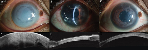
Figure 5. (a-f)- a: Slit-lamp photograph of the right eye when patient presented with a 10 days history of spontaneous hydrops shows an area of differential transparency in the mid periphery; b: Slit-lamp photograph of the right eye 1 h after the surgery shows remarkable reduction of corneal edema; c: Slit-lamp photograph on the first postoperative day shows clear visual axis; d-f: The corresponding optical coherence tomography pictures at pre-op, 1 h and 1 day are shown in d, e, and f, respectively.
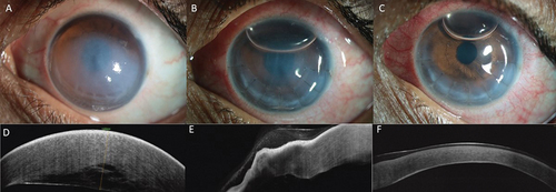
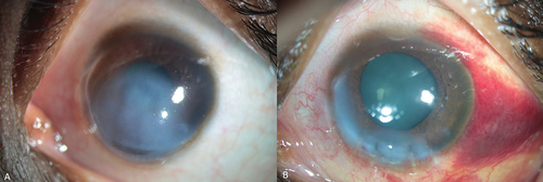
Figure 6. (a, b): a: External digital photograph of the right eye showing diffuse corneal edema; b: External photograph of the eye on the first postoperative day. (The patient was not able to cooperative for slit-lamp photographs.).
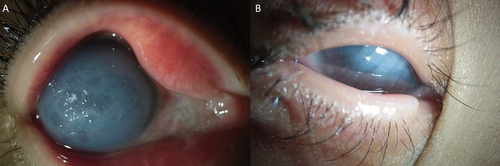
Figure 7. (a, b): a: Preoperative slit-lamp photograph of the left eye of patient 6; b: Postoperative image after 1 h of the surgery.
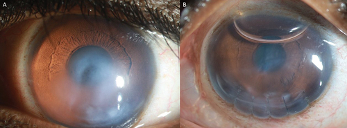
Figure 8. (a, b): a: Preoperative slit-lamp photograph of the Patient 7; b: Postoperative picture after 1 h of the surgical intervention showing marked clearing in the central cornea.