Figures & data
Table 1. Table showing interclass correlation coefficient, percentage agreement and κ value used by various authors.
Table 2. Table showing the illuminance used by various authors.
Table 3. Table showing the different ciliary body parameters, with their definitions according to various authors.
Figure 1. Figure depicting ciliary body length (CBL) and ciliary body thickness (CBT). SS: scleral spur.
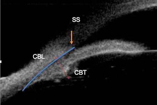
Table 4. Ciliary body thickness (CBT) and ciliary body length (CBL) in normal individuals.
Table 5. Trabecular ciliary process distance (TPCD), Trabecular ciliary angle (TCA) and Scleral ciliary process angle (SCPA).
Table 6. Significant results related to CBT and CBL of normal subjects.
Figure 2. Figure depicting ciliary muscle thickness (CMT) and ciliary process thickness (CPT). SS: scleral spur.
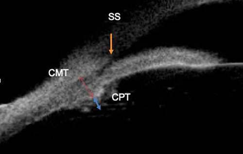
Figure 3. Figure depicting ciliary muscle thickness at the levels of CM25, CM50 and CM75, as the width of the muscle determined at points that fell 25%, 50% and 75% of the total length posterior to the scleral spur. SS:scleral spur.
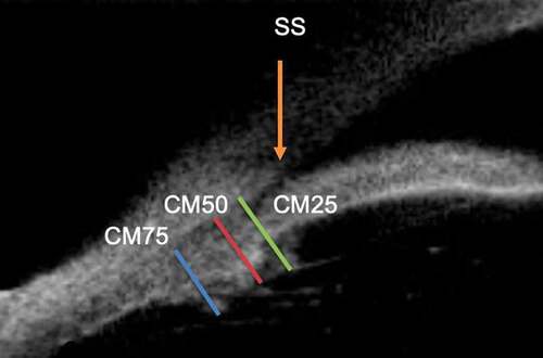
Figure 4. Figure depicting ciliary muscle length (CML) and ciliary muscle anterior length (CMAL). SS: scleral spur.
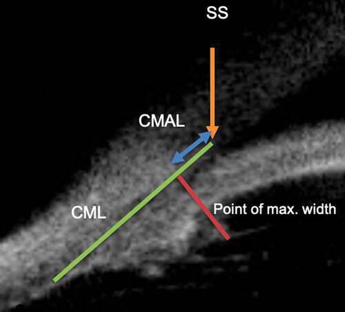
Figure 5. Figure depicting scleral ciliary process angle (SCPA) as described by Okamoto et al.Citation8 and Garudadri et al.Citation3 SS: scleral spur.
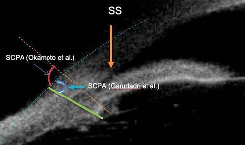
Figure 6. Figure depicting scleral ciliary process angle (SCPA), Trabecular ciliary angle (TCA), Iris ciliary angle (ICA), Trabecular ciliary process distance (TCPD) and Iris ciliary process distance (ICPD). SS: scleral spur.
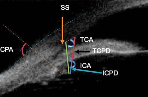
Figure 7. Figure depicting the concept of long ciliary process. A line was drawn (line C) perpendicular to the plane of the iris and this line passed through a point 750 µm anterior to the scleral spur at the corneal endothelium. A long ciliary process was defined as one whose length crossed this line, and the ciliary sulcus was said to be absent if the ciliary processes were opposed to the peripheral iris at this same location. SS: scleral spur.
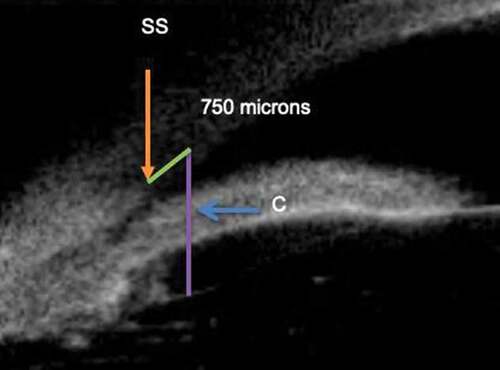
Table 7. Significant results related to angle closure.
Figure 8. Shows a uniquely thin, ill-defined and “rarefied” ciliary body, most probably owing to the stretching of the coats of the eye, with thinner ciliary body thickness (CBT) in congenital glaucoma.