
Figures & data
Figure 1. Flow diagram of open OCT data collection.
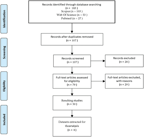
Table 1. Open OCT datasets details.
Figure 2. Comparative Samples of OCT Imaging Technologies: A. Cirrus HD OCT from OCTID, B. Topcon 3D OCT-1000 from data on OCT and fundus images, C. Spectralis OCT by Heidelberg Engineering from labeled OCT and chest X-Ray images.
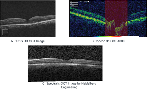
Figure 3. Distribution of patients in the OCT datasets per country. Map created adapting naturalearth_lowres’s layer using (c) 2013–2022, GeoPandas developers, an open-source python package created under the liberal terms of the BSD-3-Clause license.Citation21.
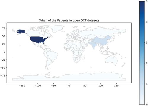
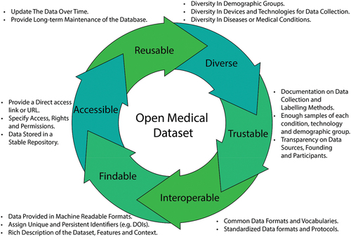
Figure 4. Key components of the ideal OCT dataset.
Supplemental material