Figures & data
Table 1. Basic data of BEN patients
Table 2. Renal size and function of BEN patients
Table 3. Renal interstitial capillaries in different stages of BEN
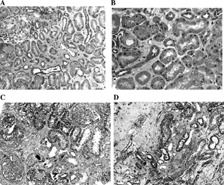
Figure 1. Varying extent of kidney cortex damage in two BEN patients. (A) Small scattered areas of discrete interstitial sclerosis with apparently normal tubuli. Segmental widening of the mesangium and discrete periglomerular sclerosis. Trichrom Masson × 100. (B) Discrete interstitial sclerosis and apparently normal proximal tubular epithelial cells on a larger magnification. A small arterial blood vessel with a thickened, partly hyalinized wall and narrowed lumen, similar to that in benign nephroangiosclerosis. Trichrom Masson × 200. (C) Larger areas of acellular interstitial sclerosis accompanied by tubular atrophy. Two glomeruli are seen: one in advanced sclerosis and the other with widening of the mesangium. Marked periglomerular sclerosis. Trichrom Masson × 100. (D) Juxtamedullary cortex with advanced acellular interstitial sclerosis and tubular atrophy. Trichrom Masson × 100.