Figures & data
Table 1 Primer sequences and annealing conditions for polymerase chain reaction
Figure 1 Effect of fenoldopam on ischemia/reperfusion induced apoptosis. Quantitative measurement of DNA fragmentation in nuclei of renal (A) cortex and (B) medulla. Each tissue section was analyzed in at least 20 areas (10 in cortex and 10 in medulla) by two blinded investigators, and the mean number of stained nuclei was calculated. The original magnification for nuclei counting was ×200. Group-wise differences were calculated using ANOVA.
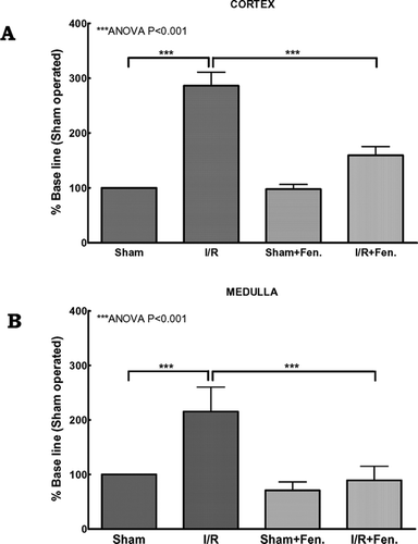
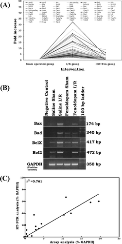
Figure 2 (A) Effect of fenoldopam on ischemia/reperfusion-induced apoptosis signal transduction genes. All 73 genes up-regulated (stringent criteria ≥ two-fold) by I/R injury were returned to the baseline level or less with fenoldopam. In each group, total RNA from six individual animals were pooled and subjected to microarray analysis. Each spot in the figure represents the expression of the particular gene in each group. A connecting line is drawn for every single gene between groups to illustrate the level of induction and attenuation for each gene after I/R injury with saline and fenoldopam, respectively. (B) RT-PCR analysis of Bax, Bad, Bcl2, and Bclx mRNA expression in rat kidney after sham operation and ischemia/reperfusion with or without fenoldopam. GAPDH was used as positive control. Negative controls without template RNA were shown in lane 1. (C) Correlation analysis of the expression levels for Bax, Bad, Bcl2, and Bclx between the microarray analysis and RT-PCR analysis.