Figures & data
Figure 1. Comparison of OGTT responses in separate groups of (A) non-diabetic and (B) streptozotocin (STZ)-treated diabetic rats treated with graded doses of FTE with control animals treated with deionized water or positive controls treated with metformin. Values are presented as means, and vertical bars indicate SE of means (n = 6 in each groups). *p < 0.01 by comparison with control animals.
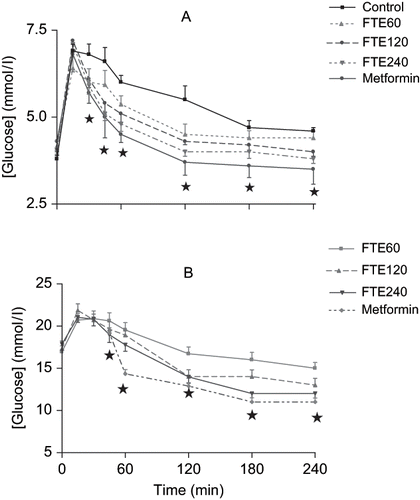
Table 1 Plasma glucose, Na+, K+, urea and creatinine concentrations and GFR in non-diabetic and STZ-diabetic control and rats administered FTE every third consecutive day for five weeks (n = 6 in all groups)
Figure 2. Effects of chronic FTE treatment on mean arterial blood pressures in (A) non-diabetic or (B) STZ-induced diabetic rats. Values are presented as means and vertical bars indicate SE of means (n = 6 in each group). *p < 0.01 by comparison with control animals.
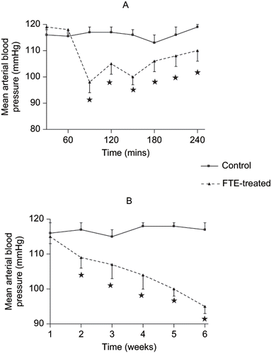
Figure 3. Viability of (A) LLC-PK1 and (B) MDBK cell lines treated with FTE. The cells were treated with 100, 200, 400, 600, 800, or 1000 μg FTE. Cell viability was determined by the Tryphan blue exclusion assay. Values for untreated control were taken as 100%. Each dose represents the mean of six treatments, while the vertical bars denote standard errors of the means.
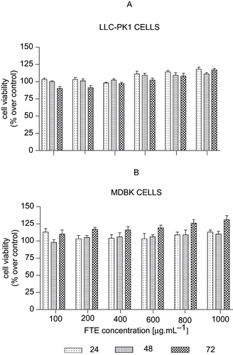
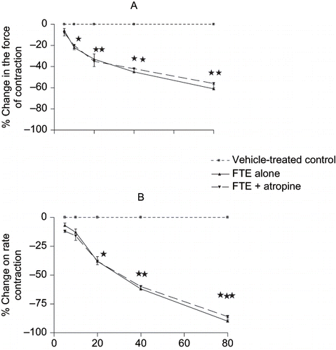
Figure 4. Effects of sequentially applied graded concen- trations of FTE (5–80 mg.mL−1) on the (A) rate and (B) force of contractions of rat isolated spontaneously beating right- and electrically driven left-atrial muscle strips, respectively. Each point represents the mean of 8–10 observations, while the vertical bars denote standard errors of the means. *p < 0.05, **p < 0.01, ***p < 0.001 versus control.