Figures & data
Figure 1. Histological examination of rat kidney, from the regions of cortex. (A, B) Control rat kidney, showing normal morphology. (C, D) Cisplatin-treated rat kidney, showing massive dilation of tubules (asterisk), tubular necrosis (arrowhead), cast formation in the lumen (arrowhead-ball), and focal mononuclear cells infiltration (arrow). (E, F) Cisplatin + vitamin C-treated rat kidney, showing slight dilation of tubules and degenerative changes (hematoxylin + eosin, scale bar: 100 μm).
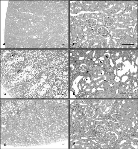
Figure 2. Photomicrographs of kidney sections stained with hematoxylin-eosin (A, D, G), Masson's trichrome (B, E, H), and periodic acid-Schiff-hemalen (C, F, I). (A, B, C) Control rats. (D, E, F) Cisplatin-treated rats, arrowhead; tubular epithelial cell exhibited enlarged nuclei with basophilic cytoplasm, arrow; thickening of the basement membrane in the renal tubules and Bowman's capsule, asterisk; enlarged periglomerular spaces. (G, H, I) Cisplatin + vitamin C-treated rats (Scale bar: 50 μm).
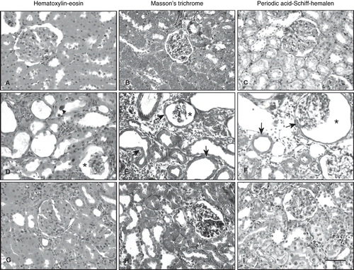
Figure 3. Electron micrographs of rat renal tubules. Left panel shows proximal renal tubules, right panel shows distal tubules. (A) Control rat. Left and right panels show normal morphology. (B) Cisplatin-treated rat. Left and right panels show numerous cytoplasmic vacuoles (v), invagination of the nuclear envelope (arrow), rounded mitochondria with disordered cristae (arrowhead), shortening and loss of basal infoldings, partial loss of cytoplasm (asterisk), and microvilli. (C) Cisplatin + vitamin C-treated rat. Left and right panels show little evidence of damaged cellular structure (uranyl acetate and lead citrate, scale bar: 1μm). Abbreviations: microvilli (Mv), nucleus (N), mitochondria (M), and basal infoldings (Bi).
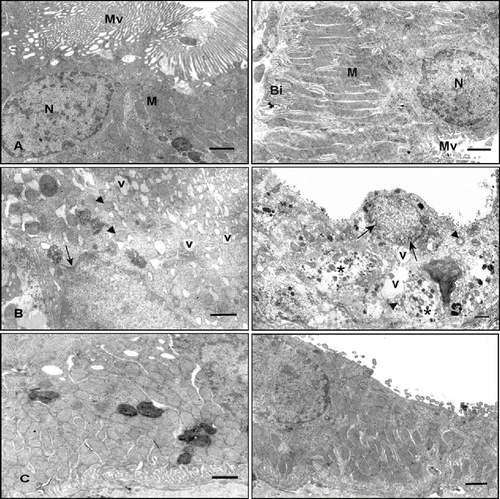
Table 1 Comparison diameter of glomerules and tubules in renal tissues