Figures & data
Table 1 Results of laboratory studies in a 43-year-old woman with acute chronic renal failure
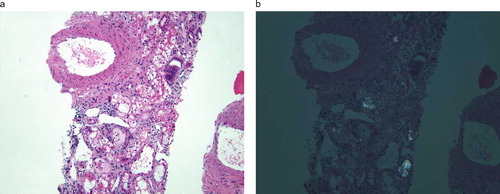
Figure 1. Kidney biopsy from a 43-year-old woman with acute chronic renal failure. The specimen is examined using an H&E stain at a magnification of 100 × the original size. In , the specimen is examined under bright-field microscopy, showing an acute tubulointerstitial process superimposed on chronic parenchymal damage. There is diffuse acute tubular injury with interstitial edema as well as substantial background fibrosis (40–50%). An active inflammatory infiltrate is seen; however, there are no granulomas and eosinophils are not prominent. In , the specimen is examined under plane-polarized illumination. White birefringence from calcium oxalate type crystals are present within nonscarred areas, suggesting recent deposition. The history of recent ethylene glycol ingestion is the likely etiology.