
Figures & data
Table 1. Laboratory data, demographic and ultrasonographic features of all patients (mean values).
Table 2. Factors affecting YM.
Figure 1. Patient 1: light microscopic images of renal tissue that stage T1 (moderate IFTA)-stage F0 (absence of IF) and their SWE image. (a) H&E ×200. (b) Masson’s trichrome ×200. (c) YM = 7.2 kPa (compatible with the stage F0 (absence of IF)).
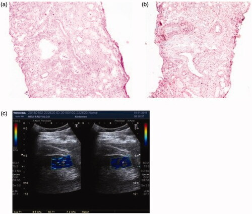
Figure 2. Patient 2: light microscopic images of renal tissue that stage T1 (moderate IFTA)-stage F2 (severe IF) and their SWE image. (a) H&E ×200. (b) Masson’s trichrome ×200. (c) YM = 29.7 kPa (compatible with the stage F2 (severe IF)).
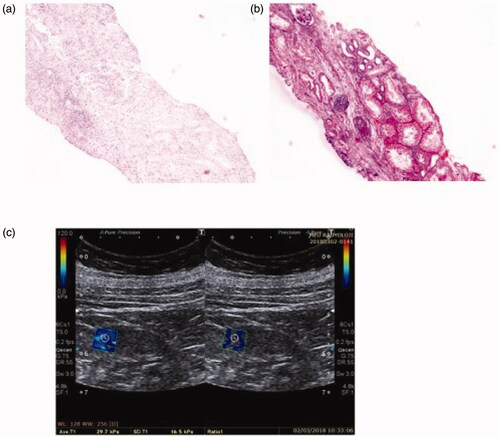
Figure 3. Patient 3: light microscopic images of renal tissue that stage T2 (severe IFTA)-stage F1 (moderate IF) and their SWE image. (a) H&E ×200. (b) Masson’s trichrome ×200. (c) YM = 21.3 kPa (compatible with the stage F1 (moderate IF)).
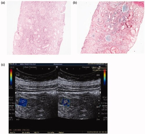
Table 3. Sensitivity, specificity, PPV, NPV, accuracy of SWE, URD, URPT, and eGFR to diagnose presence of IF.
Table 4. Sensitivity, specificity, PPV, NPV, accuracy of SWE, URD, URPT, and eGFR to diagnose presence of IFTA.