
Figures & data
Figure 1. Abdominal plain film of case 1.
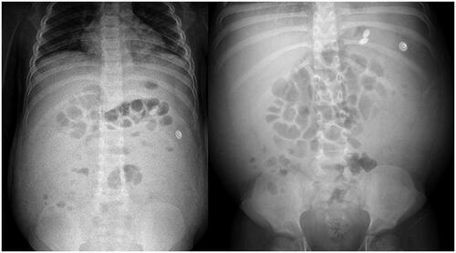
Table 1. Results of peritoneal fluid analysis and culture in 4 cases of NS with CMV peritonitis.
Figure 2. Chest radiograph of case 2.
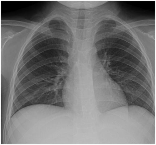
Figure 3. Renal biopsy of case 3. (A) Epithelial cells of renal tubule showed granular and vacuolar degeneration (H&E staining, ×4). (B) Mild proliferation of mesangial cells and stromal cells (H&E staining, ×400). (C & D) electron microscopy image showing minimal change disease, extensive fuzing of podocyte processes of the epithelial cells (arrow).
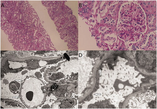
Figure 4. Chest radiograph of case 4.
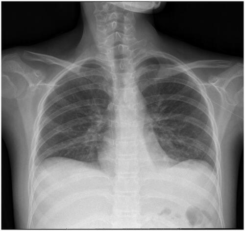
Table 2. Summary of relevant data of four patients in the literature.
Data availability statement
All meaningful data generated or analyzed in this study are included in the manuscript.