
Figures & data
Table 1. Partial biochemical indicators from May 2016 to June 2020.
Figure 1. Evolution of hemoglobin (Hb) and ferritin with anti-anemia treatment from May, 2016 to June, 2020. From May, 2016 to April, 2018, the patient had been intermittently receiving rHuEPO for renal anemia. From April, 2018 to June, 2019, the patient was treated with rHuEPO, transfusion, deferasirox and glucocorticoid for refractory anemia, iron overload, positive autoantibodies of red blood cells and EPO-antibody. However, Hb had been less than 90 g/L, even lower than 60 g/L, and ferritin had been above 2000 μg/L. In July, 2019, the patient was prescribed roxadustat for EPO-resistance renal anemia. Three months later, Hb increased to 97 g/L, and ferritin decreased to 1004 μg/L. After consumption of roxadustat for one year, Hb was 133 g/L, and ferritin was 887 μg/L. (GraphPad Prism was applied to create the picture.)
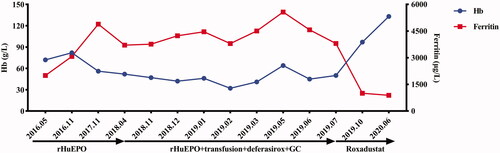
Figure 2. Fluorescence in situ hybridization of bone marrow samples for myelodysplastic syndrome. (a) 99% cells showed two green and two red signals with GLP D5S23, D5S72\EGR1 probe, indicating that the patient was without absence of 5q31. (b) 99% cells showed two green and two red signals with GLP D5S23, D5S72\CSF1R probe, indicating that the patient was without absence of 5q33. (c) 98% cells showed two green and two red signals with GLP D7S486, D7S522/CSP7 probe, indicating that the patient was without absence of chromosome 7. (d) 99% cells showed two green and two red signals with GLP D20S108/CSP8 probe, indicating that the patient was without absence of chromosome 8 and 20q12.
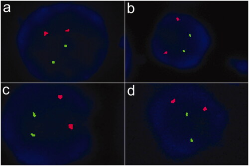
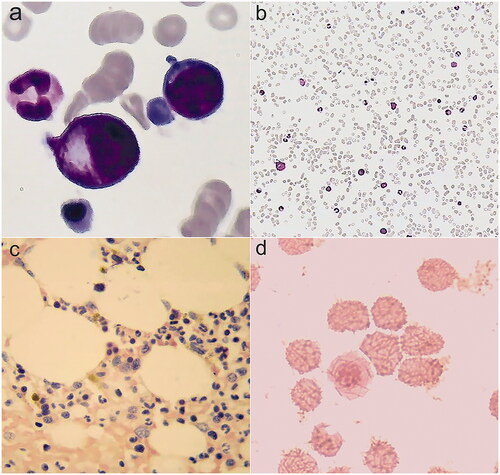
Figure 3. Bone marrow smears. (a,b) Cytologic examination in December, 2018: bone marrow was active proliferation, granulocyte was 49%, erythroid was 45%, and granulocyte: erythroid was 1.1:1. (c) Pathological examination: no pathological cells. (d) Iron stain: extracellular iron +, intracellular iron 55%, no ring sideroblasts.
Data availability
All data supporting the case are included in the manuscript.