
Figures & data
Figure 1. Pedigree of the kindred with Fabry disease. Y: years old.
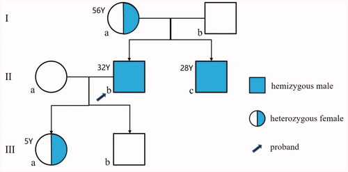
Figure 2. Skin injury. (A) Skin angiokeratoma (arrow) on the back of the proband; (B) Skin angiokeratoma (arrow) on the lower abdomen of the proband’s younger brother.
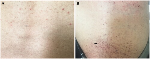
Table 1. The laboratory results of the proband.
Figure 3. (A) (HE 400×) and (B) (PAS 400×): Light microscopy showed normal glomerular volume, well-opened capillary loops, and mild segmental mesangial widening with increasing mesangial matrix. Swollen podocytes with abundant, foamy, clear cytoplasm were observed (arrow); mild chronic tubulointerstitial lesions and mild acute lesions were presented, along with flattened tubular epithelial cell and brush border loss (asterisk). (C and D) By electron microscopy, glomerular capillary loops were well-opened and basement membranes were of normal thickness. Extensive foot process fusion (black arrow) with microvillus transformation was observed in the podocyte. And osmiophilic lamellar inclusions were abundantly presented in the podocyte cytoplasm, some of which showed appearance of zebra body (white arrow).
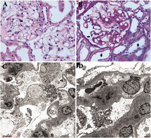
Table 2. Laboratory and clinical data of nine cases with NS in FD patients.