
Figures & data
Figure 1. Histological examination of a lymph node biopsy, fibrous tissue proliferation with numerous eosinophilic infiltration in lymph nodes.
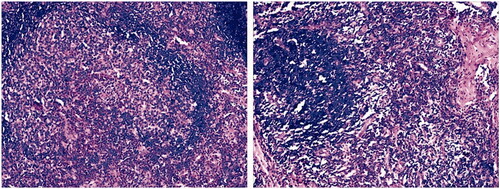
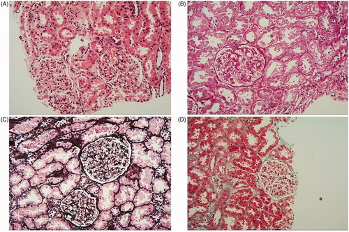
Figure 2. Histological examination of a renal biopsy showed minimal change disease (MCD) and there was no typical infiltration of eosinophil in the renal tissue. (A) The lesion shows mild in the glomerulus(H&E staining × 200). (B) Mesangial cells proliferation and mesangial matrix expansion (PAS staining × 200). (C) No thickening in glomerulus basement membrane and the glomerular capillary loops open well (PASM staining × 200). (D) There is no obvious immune complex deposition in the glomerulus (Mason staining × 200).
Data availability statement
All data supporting the case are included in the manuscript.