
Figures & data
Table 1. Clinical, pathological, and immunological features of patients with post-ESWL anti-GBM disease in our hospital.
Figure 1. Renal pathology of patient 3 with post-extracorporeal shock wave lithotripsy (ESWL) anti-GBM disease showed cellular crescent formation in a glomerulus by periodic acid-silver methenamine and Masson trichrome stain on light microscopy (400×).
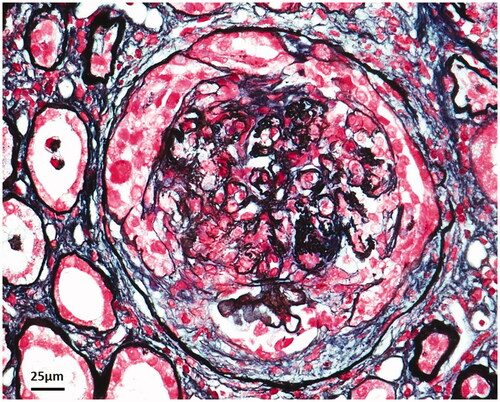
Table 2. Clinical and pathological data of previously reported post-ESWL anti-GBM disease cases.
Supplemental material