
Figures & data
Figure 1. LPS triggered pyroptotic cell death and inflammatory responses in HK-2 cells. (A) Cell proliferation and (B) viability were examined by MTT assay and trypan blue staining assay. (C,D) The HK-2 cells were stained with Annexin V-FITC and PI, and the apoptotic cell ratio was determined by flow cytometer. (E,F) The pyroptosis-associated biomarkers in HK-2 cells were measured by Western Blot analysis. The uncropped WB images are shown in Figure S1. (G) Real-time qPCR and (H) ELISA were performed to evaluate inflammatory cytokines generation and secretion in HK-2 cells and its supernatants. Individual experiment had three times of biological repetitions, and each biological replicate contained three times of technical replicates, and *p < .05.
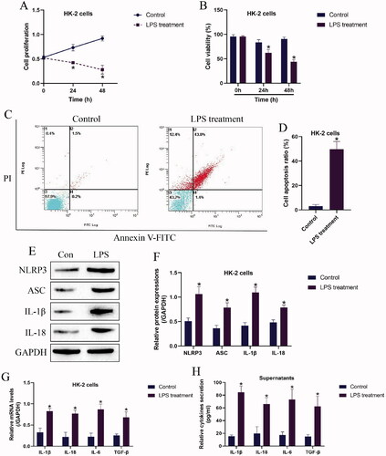
Figure 2. The regulating effects of LPS treatment on the LncRNA MALAT1/microRNA-135b-5p axis. The expression levels of (A) LncRNA MALAT1 and (B) microRNA-135b-5p were detected by Real-Time qPCR. (C) Prediction and (D) validation of the binding sites in LncRNA MALAT1 and microRNA-135b-5p. (E,F) The LncRNA MALAT1/microRNA-135b-5p axis was manipulated in HK-2 cells, and the vectors transfection efficacy was examined by Real-Time qPCR. (G–I) LPS promoted NLRP3 expressions in HK-2 cells by regulating the LncRNA MALAT1/microRNA-135b-5p axis. The uncropped WB images are shown in Figure S2. Individual experiment had three times of biological repetitions, and each biological replicate contained three times of technical replicates, and *p < .05.
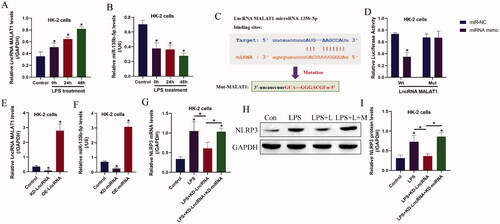
Figure 3. LPS triggered inflammatory cell death in HK-2 cells by modulating LncRNA MALAT1 and microRNA-135b-5p. Examination of (A) cell proliferation and (B) viability by MTT assay and trypan blue staining assay. (C,D) Annexin V-FITC and PI double staining assay was utilized to determine cell apoptosis ratio in HK-2 cells. (E,F) Western Blot analysis was performed to measure the expression levels of NLRP3, ASC, IL-1β, and IL-18 in HK-2 cells. The uncropped WB images are shown in Figure S3. (G) Real-Time qPCR and (H) ELISA were respectively performed to examine production and secretion of the inflammation-associated cytokines in HK-2 cells and its supernatants. Individual experiment had three times of biological repetitions, and each biological replicate contained three times of technical replicates, and *p < .05.
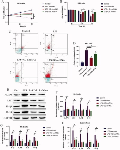
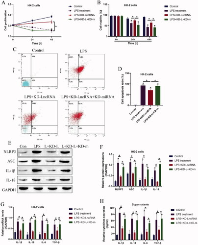
Figure 4. Knock-down of LncRNA MALAT1 attenuated LPS-induced cell pyroptosis in HK-2 cells by releasing microRNA-135b-5p. (A) Cell proliferation and (B) viability were determined by MTT assay and trypan blue staining assay. (C,D) Cell apoptosis ratio in HK-2 cells was examined. (E,F) Western Blot analysis was performed to examine the levels of pyroptosis-associated proteins. The uncropped WB images are shown in Figure S4. (G,H) The levels of inflammation-associated cytokines were determined in HK-2 cells and its supernatants. Individual experiment had three times of biological repetitions, and each biological replicate contained three times of technical replicates, and *p < .05.