Figures & data
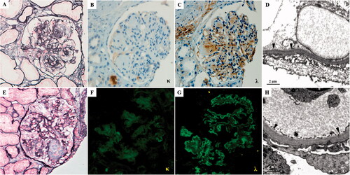
Figure 1. The pathological findings of the first (A–D) and second (E–H) renal biopsy. (A) The glomeruli exhibit sclerotic mesangial nodules with severe mesangial hypercellularity (PASM, ×400). (B) Kappa light chain is negative by immunohistochemistry staining on paraffin tissue (×400). (C) Lambda light chain deposit in nodular sclerosis area and along capillary wall and TBM by immunohistochemistry staining on paraffin tissue (×400). (D) Electron microscopy of formalin-fixed renal tissue. The granular dense (arrowhead) deposits along the inner side of GBM (×12,000). (E) The nodular sclerosis was significantly reduced than the first biopsy (PASM, ×400). (F) The kappa-light chain stains negatively. (G) Immunofluorescence staining showed that the lambda-light chain is positive along the capillary wall of glomeruli (×400). (H) Electron microscopy. There is trace granular (arrowhead) dense deposition along the inner side of GBM (×12,000).