
Figures & data
Table 1. Demographic and clinical evaluation of patients with C3GN and monoclonal gammopathy.
Table 2. Complement and hematological evaluation and kidney biopsy findings of patients with C3GN and monoclonal gammopathy.
Figure 1. Light, immunofluorescence, and electron microscopy findings in a C3GN patient with monoclonal gammopathy (patient #15). (A) Light microscopy showed a membranoproliferative pattern of injury with a small fibrocellular crescent formation (Periodic acid-silver methenamine + Masson trichrome staining, ×400). (B) Immunofluorescence staining showed bright C3 in the mesangial and along segmental capillary walls (×400). (C) Electron microscopy showed electron-dense deposits in subendothelial, intramembranous and mesangial regions (×8000).
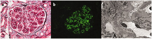
Figure 2. Light, immunofluorescence, and electron microscopy findings in a C3GN patient with TMA-like lesions (patient #17). (A) Light microscopy showed a mesangial proliferative pattern of injury with endocapillary hypercellularity and segmental sclerosis (Periodic acid-silver methenamine + Masson trichrome staining, B × 400). (B) Immunofluorescence studies showed bright C3 in the mesangial and along segmental capillary walls (×400). (C): Electron microscopy showed electron-dense deposits in mesangial and intramembranous regions and subendothelial edema with narrowing of the capillary lumen (×8000).
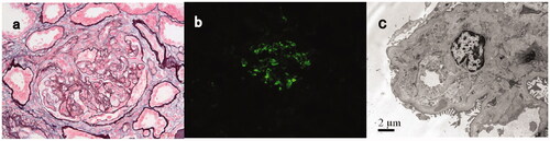
Table 3. Treatment and kidney measures at follow up of patients with C3GN and monoclonal gammopathy.
Table 4. Previous reports of patients with C3G and monoclonal gammapathy.
Data availability statement
All data generated or analyzed during this study are included in this published article.