
Figures & data
Figure 1. A large number of purple granular inclusions and needle-shaped crystals could be seen in the cytoplasm of bone marrow plasma cells (Wright Giemsa stain, 1000 × original magnification).
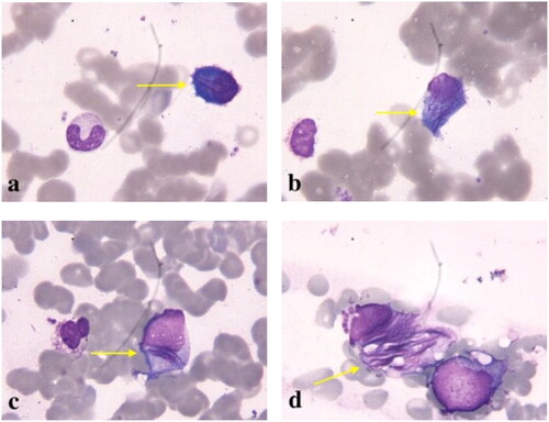
Figure 2. Crystalline inclusions in podocytes (a: HE; b: PAS), proximal tubular epithelial cells (c, d: PAS) and histiocytes (e: PAS; f: HE) (original magnification all ×1000).
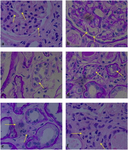
Figure 3. Periodic acid–Schiff (PAS)-positive, heterogeneous, and granular casts could be seen in the tubular lumens (a: PAS, original magnification × 100; b: PAS, original magnification × 200; c: PAS, original magnification × 400).
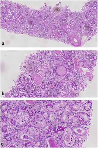
Figure 4. On immunohistochemistry, kappa light chain-restricted expression (a: κ, 200 × original magnification) while lambda was negative (b: λ, 200 × original magnification) in proximal tubular epithelial cells (c: κ, 400 × original magnification), histiocytes (d: κ, 400 × original magnification), and podocytes (e: κ, 400 × original magnification).
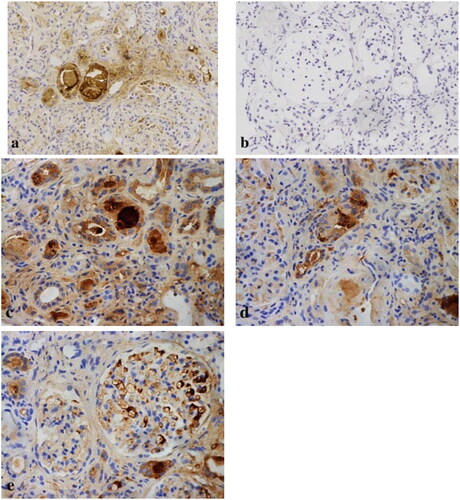
Figure 5. Immunohistochemical stains for immunoglobulins confirm the nature of the crystalline inclusions within the histiocytes. Immunoreactivity for kappa light chain is strong within the histiocytes as well as in the plasma cells, which demonstrates kappa light chain restriction. Immunohistochemistry showed that there were histiocytosis in renal interstitium (a: CD68, 100 × original magnification; b: κ, 100 × original magnification; c: CD68, 400 × original magnification).
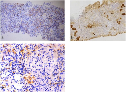
Figure 6. Immunofluorescence with pronase digestion on formalin-fixed paraffin-embedded tissue shows bright staining for kappa (400 × original magnification).
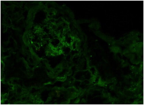
Figure 7. Electron microscopy showed crystalline inclusions in the podocytes (a, b), mesangial cells (c, d), proximal tubular epithelial cells (e, f), and histiocytes (g, h) (uranyl acetate and lead citrate staining, original magnification: a, f, g: ×4, 200, bar = 2 μm; b: ×6, 000, bar = 2 μm; c: ×9, 900, bar = 1 μm; d: ×20, 500, bar = 500 nm; e: ×11, 500, bar = 1 μm; h: ×6, 000, bar = 2 μm).
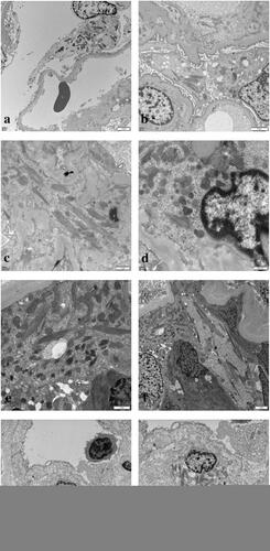
Figure 8. Immunoelectron microscopy showed that kappa light chain was strongly positive (a, b, c) in the crystalline inclusions and lysosomes of proximal tubular epithelial cells and podocytes, while lambda light chain was negative (d) (original magnification: a, b: κ × 26, 500, bar = 200 nm; c: κ × 11, 500, bar = 1 μm; d: λ × 20, 500, bar = 500 nm).
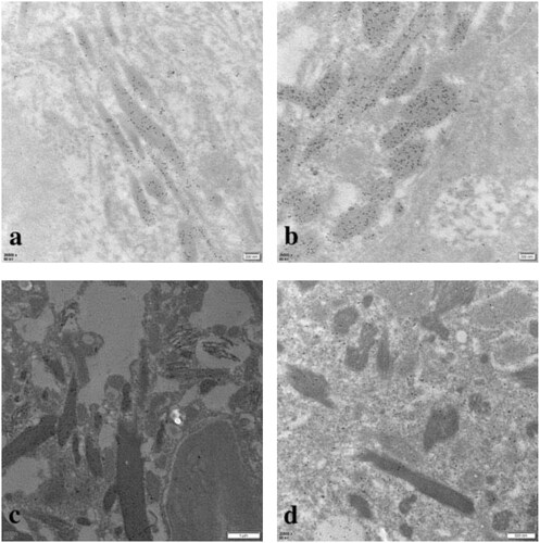
Table 1. Overview of CSH with renal involvement.
Data availability statement
All data generated or analyzed during this study are included in this published article.