2,746
Views
2
CrossRef citations to date
0
Altmetric
State-of-the-Art Review

Advances in imaging techniques to assess kidney fibrosis

, , &
Article: 2171887 | Received 19 May 2022, Accepted 13 Jan 2023, Published online: 01 Feb 2023

Figures & data

Figure 1. The pathological foundation of imaging techniques to assess kidney fibrosis. BOLD-MRI: Blood oxygenation level-dependent MRI; ASL-MRI: Arterial spin labeling MRI; SE: strain elastography; SWE: shear wave elastography; MRE: magnetic resonance elastography; PAI: photoacoustic imaging; ECM: extracellular matrix; MTI: magnetization transfer imaging; DWI: diffusion weighted imaging; DTI: diffusion tensor imaging; IVIM: intravoxel incoherent motion imaging; DKI: diffusion kurtosis imaging. The increase and deposition of ECM lead to the increase of kidney stiffness and macromolecule content, meanwhile, decrease the tissue oxygenation and perfusion, restrict the diffusion of water molecules. This pathologic change is the foundation of different imaging techniques to assess kidney fibrosis.

Figure 1. The pathological foundation of imaging techniques to assess kidney fibrosis. BOLD-MRI: Blood oxygenation level-dependent MRI; ASL-MRI: Arterial spin labeling MRI; SE: strain elastography; SWE: shear wave elastography; MRE: magnetic resonance elastography; PAI: photoacoustic imaging; ECM: extracellular matrix; MTI: magnetization transfer imaging; DWI: diffusion weighted imaging; DTI: diffusion tensor imaging; IVIM: intravoxel incoherent motion imaging; DKI: diffusion kurtosis imaging. The increase and deposition of ECM lead to the increase of kidney stiffness and macromolecule content, meanwhile, decrease the tissue oxygenation and perfusion, restrict the diffusion of water molecules. This pathologic change is the foundation of different imaging techniques to assess kidney fibrosis.

Table 1. Features of noninvasive imaging techniques for assessing kidney fibrosis.
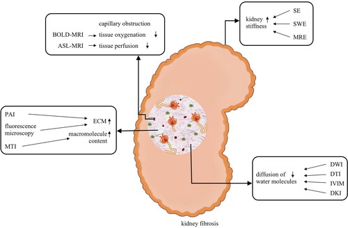
Table 2. Studies on noninvasive imaging techniques for assessing kidney fibrosis.