Figures & data
Table 1. The primer sequences used in this study.
Figure 1. MALAT1 and miR-30c were differentially expressed in calcified VSMCs. (A) Alizarin red S staining indicated more mineralized nodules in the Cal-medium group than the DMEM group; (B) calcium assay showed higher calcium content in the Cal-medium group; (C) Western blot revealed higher expression of Runx2 and lower expression of α-SMA in the Cal-medium group; (D) qRT-PCR results showed that MALAT1 was increased and miR-30c was decreased in the Cal-medium group. MALAT1: metastasis-associated lung adenocarcinoma transcript 1; VSMCs: vascular smooth muscle cells; Cal-medium: calcifying medium; DMEM: Dulbecco’s Modified Eagle Medium; Runx2: runt-related transcription factor 2; α-SMA: alpha smooth muscle actin; qRT-PCR: quantitative reverse transcription PCR. N = 3. At least two independent experiments were performed. *p < 0.05 vs. the DMEM group.
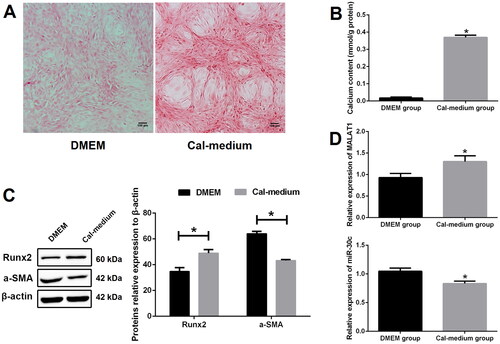
Figure 2. Knockdown of MALAT1 greatly suppressed VSMCs calcification. (A) qRT-PCR showed that the expression of MALAT1 was significantly decreased after MALAT1 shRNA transfection; (B) MALAT1 shRNA transfection reduced mineralized nodules as indicated by Alizarin red S staining; (C) MALAT1 shRNA transfection reduced cell calcium content as indicated by calcium assay; (D) Western blot demonstrated lower expression of Runx2 and higher expression of α-SMA after MALAT1 shRNA transfection. MALAT1: metastasis-associated lung adenocarcinoma transcript 1; VSMCs: vascular smooth muscle cells; qRT-PCR: quantitative reverse transcription PCR; NC shRNA: negative control short hairpin RNA; Runx2: runt-related transcription factor 2; α-SMA: alpha smooth muscle actin. N = 3. At least two independent experiments were performed. *p < 0.05 vs. the NC shRNA group.
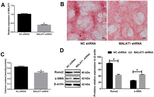
Figure 3. Up-regulation of MALAT1 promoted VSMCs calcification. (A) qRT-PCR showed that the expression of MALAT1 was significantly up-regulated after Lv-MALAT1 transfection; (B) Alizarin red S staining indicated more mineralized nodules in the Lv-MALAT1 group; (C) calcium assay demonstrated higher calcium content in the Lv-MALAT1 group; (D) Western blot demonstrated higher expression of Runx2 and lower expression of α-SMA in the Lv-MALAT1 group. MALAT1: metastasis-associated lung adenocarcinoma transcript 1; VSMCs: vascular smooth muscle cells; qRT-PCR: quantitative reverse transcription PCR; Lv-NC: negative control lentivirus; Lv-MALAT1: MALAT1 lentivirus; Runx2: runt-related transcription factor 2; α-SMA: alpha smooth muscle actin. N = 3. At least two independent experiments were performed. *p < 0.05 vs. the Lv-NC group.
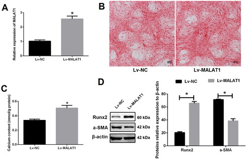
Figure 4. MALAT1 directly sponged miR-30c. (A) putative binding sites of MALAT1 with miR-30c as predicted by the ENCORI platform; (B) qRT-PCR showed that miR-30c expression was increased after MALAT1 knockdown and decreased after MALAT1 up-regulation; (C) Dual-luciferase reporter assay indicated that miR-30c mimics could significantly reduce the luciferase activity of cells transfected with MALAT1-WT; (D) qRT-PCR showed that miR-30c expression was increased after miR-30c mimics transfection; (E and F) up-regulation of miR-30c with mimics promoted VSMCs calcification as indicated by Alizarin red S staining and calcium assay. MALAT1: metastasis-associated lung adenocarcinoma transcript 1; qRT-PCR: quantitative reverse transcription PCR; NC shRNA: negative control short hairpin RNA; Lv-NC: negative control lentivirus; Lv-MALAT1: MALAT1 lentivirus. N = 3. At least two independent experiments were performed. *p < 0.05 vs. NC groups or cells cotransfected with MALAT1-WT and NC mimics.
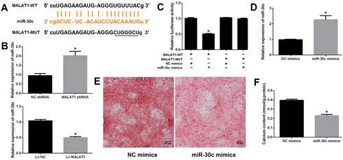
Figure 5. Runx2 is a direct target of miR-30c. (A) miR-30c putative target site in the 3’-untranslated region of Runx2 as predicted by the ENCORI platform; (B and C) qRT-PCR and western blot showed that Runx2 expression was decreased after miR-30c mimics transfection; (D) dual-luciferase reporter assay indicated that miR-30c mimics could significantly reduce the luciferase activity of cells transfected with Runx2-WT. MALAT1: metastasis-associated lung adenocarcinoma transcript 1; Runx2: runt-related transcription factor 2; qRT-PCR: quantitative reverse transcription PCR. N = 3. At least two independent experiments were performed. *p < 0.05 vs. NC groups or cells cotransfected with Runx2-WT and NC mimics.
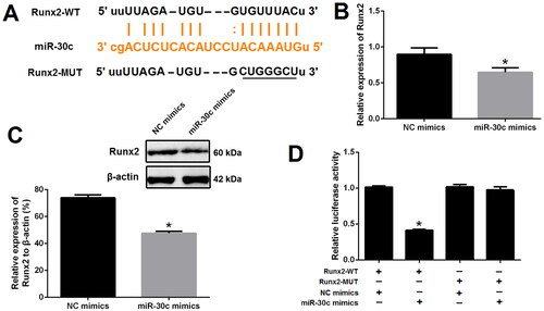
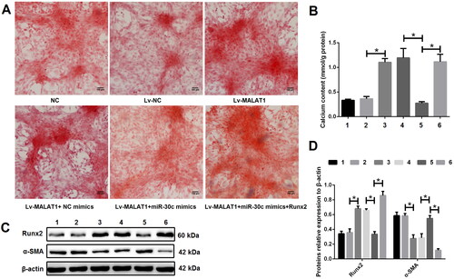
Figure 6. The effect of MALAT1 on VSMCs calcification was at least partially through regulating the miR-30c/Runx2 axis. (A) Representative Alizarin red S staining pictures; (B) calcium assay showed that the Lv-MALAT1 vector induced higher calcium content, which was decreased after adding miR-30c mimics but increased again after adding Runx2 over-expression vector; (C and D) Western blot results showed that Lv-MALAT1 vector increased Runx2 but decreased α-SMA expression, this effect was reversed after adding miR-30c mimics but reversed again after adding Runx2 over-expression vector. 1 represent the NC group, 2 represent the Lv-NC group, 3 represent the Lv-MALAT1 group, 4 represent the Lv-MALAT1 + NC mimic group, 5 represent the Lv-MALAT1 + miR-30c group and 6 represent the Lv-MALAT1+ miR-30c mimic + Runx2 group. NC: negative control; Lv-NC: negative control lentivirus; Lv-MALAT1: MALAT1 lentivirus; Runx2: runt-related transcription factor 2; α-SMA: alpha smooth muscle actin. N = 3. At least two independent experiments were performed. *p < 0.05.