1,213
Views
0
CrossRef citations to date
0
Altmetric
Brief Report

Zero-contrast imaging for the assessment of transcatheter aortic valve implantation in candidates with renal dysfunction

ORCID Icon, ORCID Icon, ORCID Icon, ORCID Icon, ORCID Icon, , , , ORCID Icon, ORCID Icon & ORCID Icon show all
Article: 2224888 | Received 06 Mar 2023, Accepted 08 Jun 2023, Published online: 23 Jun 2023

Figures & data

Figure 1. Multimodality imaging for the assessment of TAVI candidates with renal dysfunction. Femoro-iliac-aortic endovascular access (A) was evaluated by duplex scan ultrasound (upper panel showing the left and right femoral arteries), computed tomography (arrowheads in the mid-upper panel indicate the left and right femoral arteries), and nuclear magnetic resonance imaging (arrowheads in the mid-upper panel indicate the left and right femoral arteries). The aortic root and aortic annulus (B) were evaluated using computed tomography (upper panel), nuclear magnetic resonance imaging (red asterisk in the middle panel indicates the aortic annulus region), and echocardiography (lower panel), which were reassessed for confirmation during the procedure (C, left panel). The final shape of the metallic frame of the prosthesis is shown in the right panel of panel C.

Figure 1. Multimodality imaging for the assessment of TAVI candidates with renal dysfunction. Femoro-iliac-aortic endovascular access (A) was evaluated by duplex scan ultrasound (upper panel showing the left and right femoral arteries), computed tomography (arrowheads in the mid-upper panel indicate the left and right femoral arteries), and nuclear magnetic resonance imaging (arrowheads in the mid-upper panel indicate the left and right femoral arteries). The aortic root and aortic annulus (B) were evaluated using computed tomography (upper panel), nuclear magnetic resonance imaging (red asterisk in the middle panel indicates the aortic annulus region), and echocardiography (lower panel), which were reassessed for confirmation during the procedure (C, left panel). The final shape of the metallic frame of the prosthesis is shown in the right panel of panel C.

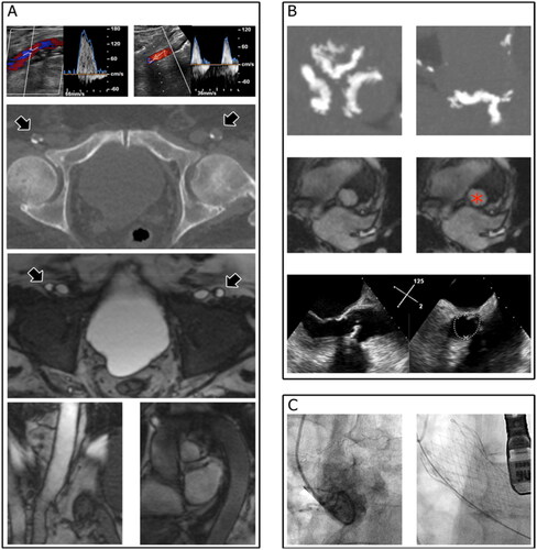
Table 1. Patients characteristics.

Table 2. Main findings of multi-modality imaging.

Table 3. Procedure characteristics.

Table 4. Renal function and in-hospital clinical outcomes.