
Figures & data
Figure 1. Progression of the cutaneous lesion (the metatarsal bone, the third toe, and the little toe suffered the most and presented with ulcers within one week, accompanied by pain and progressive cutaneous darkening and sclerosis. The skin temperature was low at the initial stage. In addition, the patient had weakened bilateral dorsalis pedis pulses, especially on the right side).
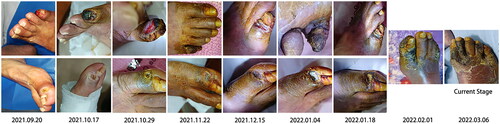
Figure 2. Echocardiography manifestations [A-B: the aortic and mitral mechanical valves (red arrows) were well placed with good activity of the valve leaflets and normal perivalvular surroundings with iso-echogenicity attached. C: There was no significant thickening of the tricuspid valve leaflet. Further, the valve showed good opening but poor closing].
![Figure 2. Echocardiography manifestations [A-B: the aortic and mitral mechanical valves (red arrows) were well placed with good activity of the valve leaflets and normal perivalvular surroundings with iso-echogenicity attached. C: There was no significant thickening of the tricuspid valve leaflet. Further, the valve showed good opening but poor closing].](/cms/asset/f2527c28-cee6-44e9-94fd-0ede2cd9fc10/irnf_a_2264401_f0002_c.jpg)
Figure 3. Vascular ultrasound of the lower extremities (there was no evident abnormality in the diameter of the bilateral lower limb artery. However, the walls were rough with multiple hyperechoic and hypoechoic plaques. Extensive calcification of the arterial media was detected. A: right femoral artery; B: right posterior tibial artery; C: right dorsalis pedis artery; red arrows show calcification areas of the arterial media).
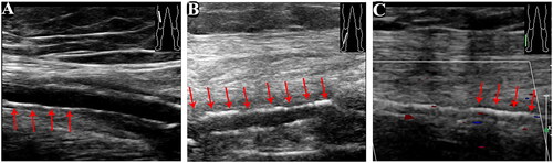
Table 1. A Summary of reported cases of calciphylaxis and prosthetic cardiac valve replacement treated with warfarin.