
Figures & data
Figure 1. Chemical structure of oridonin.
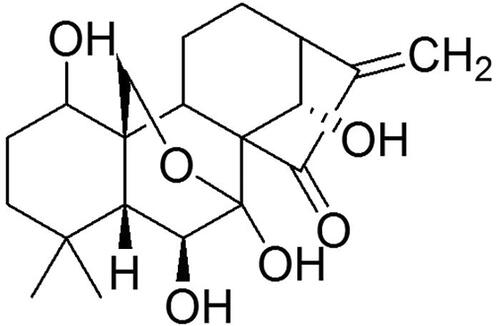
Table 1. Primers used for reverse transcription-quantitative PCR.
Figure 2. Effects of Ori on general changes and biochemical indices in diabetic rats.
(A) Effect of Ori on blood glucose. (B–D) Effect of Ori on BUN, Scr and 24-h urinary protein concentrations. **p < 0.01 vs. the NC group; ***p < 0.001 vs. the NC group; ###p < 0.001 vs. the DN group. Ori: oridonin; BUN: blood urea nitrogen; Scr: serum creatinine; NC: normal control; DN: diabetic nephropathy; DN + Ori: diabetic nephropathy + oridonin.
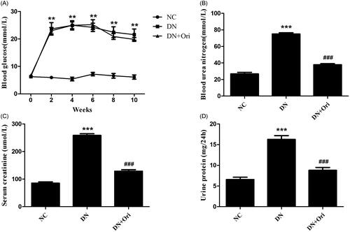
Figure 3. Effects of Ori on renal pathological changes in diabetic rats.
PAS and MT staining of kidney tissues. **p < 0.01 vs. the NC group, ###p < 0.001 vs. the DN group. PAS: periodic acid-Schiff; MT: Masson’s trichrome; NC: normal control; DN: diabetic nephropathy; DN + Ori: diabetic nephropathy + oridonin.
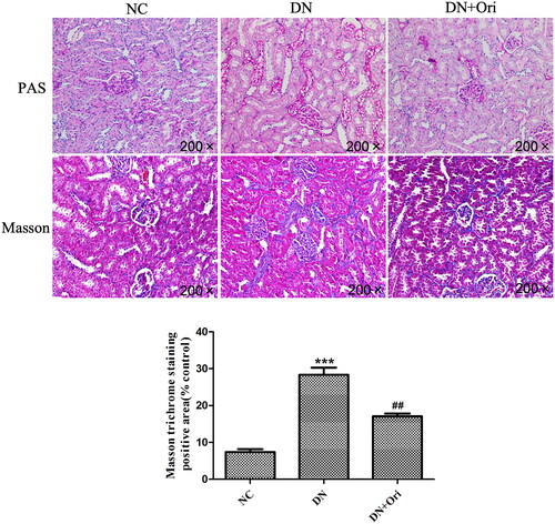
Figure 4. Effects of Ori on renal fibrosis in diabetic rats.
Immunohistochemical analysis of Col I and FN expression in kidney tissues. ***p < 0.001 vs. the NC group, ###p < 0.001 vs. the DN group. Col I, collagen I; FN: fibronectin; NC: normal control; DN: diabetic nephropathy; DN + Ori: diabetic nephropathy + oridonin.
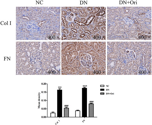
Figure 5. Expression of the Wnt/β-catenin signaling pathway in the kidneys of diabetic rats.
(A) Protein expression levels of Wnt4, GSK3β, p-GSK3β, β-catenin and α-SMA in rat kidney tissues. (B) qPCR analysis of the mRNA expression of Wnt4, GSK3β, β-catenin and α-SMA in rat kidney tissues. **p < 0.01 vs. the NC group; #p < 0.05 vs. the DN group; ##p < 0.01 vs. the DN group; ###p < 0.001 vs. the DN group. p-GSK3β: phospho-glycogen synthase kinase-3β; α-SMA: α-smooth muscle actin; qPCR: quantitative polymerase chain reaction.
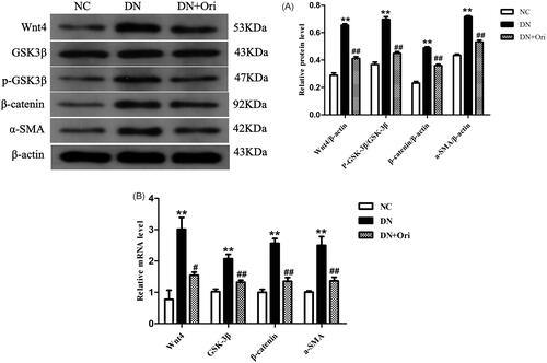
Figure 6. Effects of Ori on cell viability.
(A) HK-2 cells were treated with different concentrations of HG (0, 10, 20, or 30 mM) for 48 h, after which their viability was detected via CCK-8 assays. (B) HK-2 cells were treated with HG (30 mM) for different durations (0, 12, 24, and 48 h), and their viability was detected by CCK-8 assays. (C) HK-2 cells were pretreated with different concentrations of Ori (2.5, 5, or 10 μM) for 2 h and then treated with 30 mM HG for 48 h, after which their viability was detected via CCK-8 assays. **p < 0.01 vs. the NG group; ***p < 0.001 vs. the NG group; #p < 0.05 vs. the HG group; ##p < 0.01 vs. the HG group; HG: high glucose; CCK-8: Cell Counting Kit-8; NG: normal glucose control (5.5 mmol/L glucose); MG: mannitol control (19.5 mmol/L mannitol + 5.5 mmol/L glucose); NG + Ori: 5.5 mmol/L glucose + 5 μmol/L oridonin; HG: 30.0 mmol/L glucose; HG + Ori: 30.0 mmol/L glucose + 5 μmol/L oridonin.
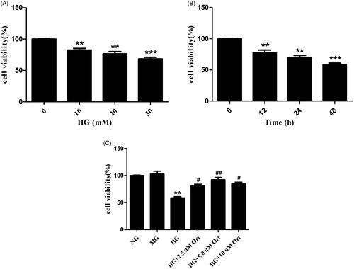
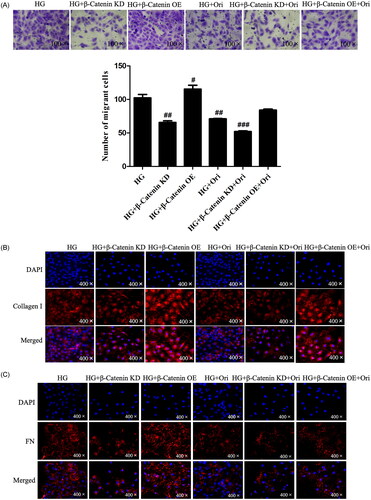
Figure 7. Ori inhibited fibrosis in HG-treated HK-2 cells.
(A) Transwell assays were used for the assessment of cell migration. (B & C) Col I and FN were detected by specific immunofluorescence staining. (D) Quantification of the immunofluorescence staining of Col I and FN. **p < 0.01 vs. the NG group; #p < 0.05 vs. the HG group; ##p < 0.01 vs. the HG group; Col I: collagen I; FN: fibronectin; HG: high glucose; NG: normal glucose control (5.5 mmol/L glucose); MG: mannitol control (19.5 mmol/L mannitol + 5.5 mmol/L glucose); NG + Ori: 5.5 mmol/L glucose + 5 μmol/L oridonin; HG: 30.0 mmol/L glucose; HG + Ori: 30.0 mmol/L glucose + 5 μmol/L oridonin.
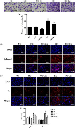
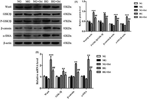
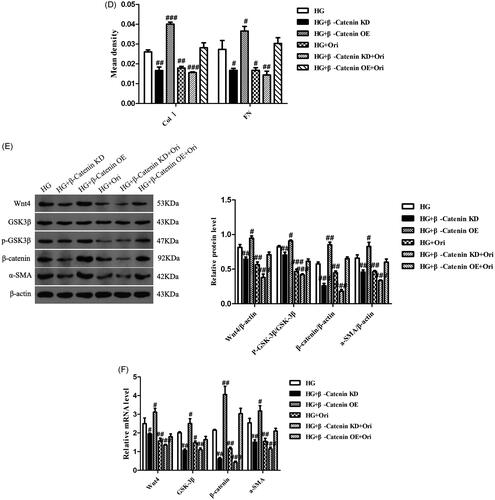
Figure 8. Expression of proteins in the Wnt/β-catenin signaling pathway in HG-treated HK-2 cells.
(A) Protein expression levels of Wnt4, GSK3β, p-GSK3β, β-catenin and α-SMA in HK-2 cells. (B) qPCR analysis of the mRNA expression of Wnt4, GSK3β, β-catenin and α-SMA in HK-2 cells. **p < 0.01 vs. the NC or NG group, ***p < 0.001 vs. the NC or NG group, #p < 0.05 vs. the DN or HG group, ##p < 0.01 vs. the DN or HG group, and ###p < 0.001 vs. the DN or HG group. p-GSK3β, phospho-glycogen synthase kinase-3β; α-SMA: α-smooth muscle actin; qPCR: quantitative polymerase chain reaction.
Data availability statement
The data that support the findings of this study are available from the corresponding author upon reasonable request.