
Figures & data
Figure 1. Study selection flowchart.
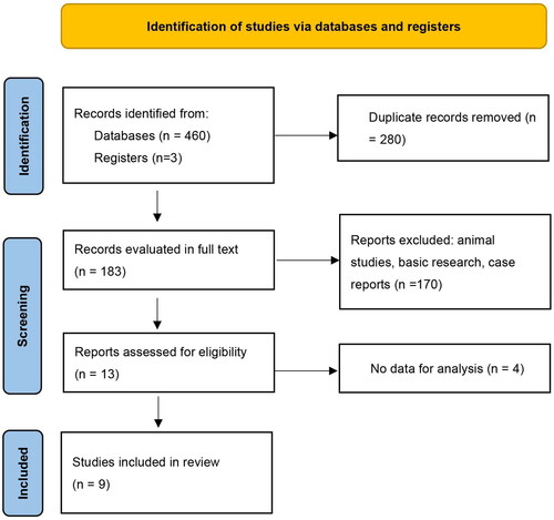
Figure 2. Forest plot of malignancy prevalence in THSD7A-associated MN.
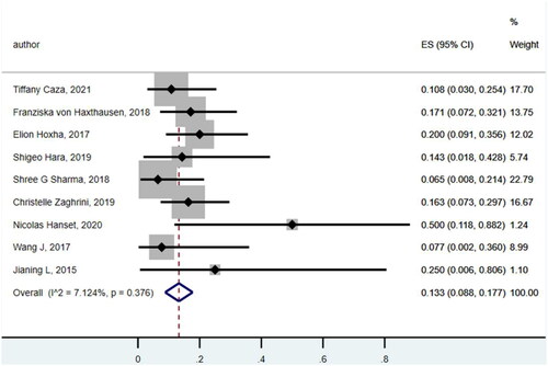
Figure 3. Funnel plot of including studies.
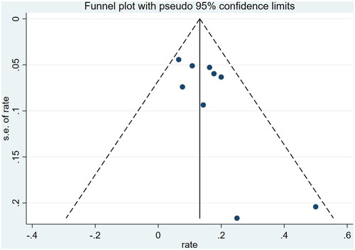
Figure 4. Representative images of glomerular and tumor staining for THSD7A. (A) Patient no. 13 with positive glomerular THSD7A staining. (B) Patient no. 14 with positive glomerular THSD7A staining. (C) Patient no. 15 with positive glomerular THSD7A staining. (D) Patient no. 14 with positive THSD7A staining of small cell lung cancer tissue (immunohistochemistry staining, magnification ×400).
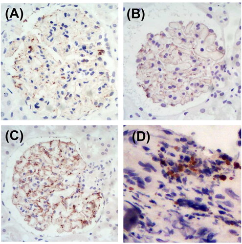
Table A1. Quality evaluation of each study using the Newcastle-Ottawa Quality Assessment Scale.
Table 1. Literature review of malignancy in THSD7A-associated MN.
Table 2. Clinical manifestations and prognosis of 15 patients with THSD7A-associated MN in our center.
Table 3. Immunofluorescence deposition of renal tissue in 15 patients with THSD7A-associated MN.
Table 4. Serum antibodies and antigen staining of renal tissues in 15 cases of THSD7A-associated MN.
Data availability statement
The data that support the findings of this study are available from the corresponding author upon reasonable request.