Publication Cover
CRANIO®
The Journal of Craniomandibular & Sleep Practice
Volume 35, 2017 - Issue 2
7,344
Views
6
CrossRef citations to date
0
Altmetric
Special report

Airway Centric® TMJ philosophy/Airway Centric® orthodontics ushers in the post-retraction world of orthodontics

(DDS, MSD) & (DDS, MS)

Figures & data

Figure 1 The patient illustrated above presented with a Class II Division 1 malocclusion and was treated with Orthotropics® to advance the maxillary anterior teeth, followed by advancement of the mandible. The airway improved dramatically in her case as both jaws were developed forward.
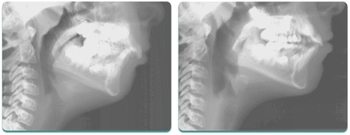
Figure 1 The patient illustrated above presented with a Class II Division 1 malocclusion and was treated with Orthotropics® to advance the maxillary anterior teeth, followed by advancement of the mandible. The airway improved dramatically in her case as both jaws were developed forward.

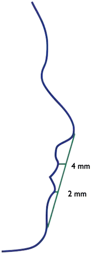
Figure 2 Esthetic line.

Figure 2 Esthetic line.

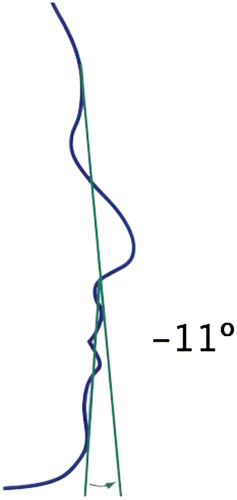
Figure 3 Facial contour angle.

Figure 3 Facial contour angle.

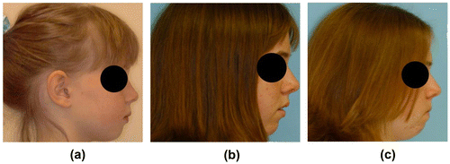
Figure 4 This little girl’s face grew more vertically (less forward) due to poor rest oral posture.

Figure 4 This little girl’s face grew more vertically (less forward) due to poor rest oral posture.

Figure 5 Patient presented with generalized spacing of the upper arch in A. Spacing was closed in the anterior and placed between the upper second bicuspid teeth and first molar teeth without retracting or reducing the arch circumference in B.
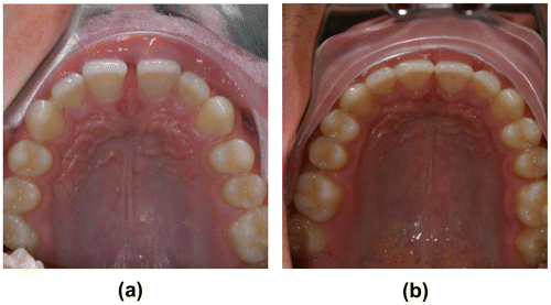
Figure 5 Patient presented with generalized spacing of the upper arch in A. Spacing was closed in the anterior and placed between the upper second bicuspid teeth and first molar teeth without retracting or reducing the arch circumference in B.

Figure 6 The patient pictured above had his snoring completely resolve after we advanced his lower anterior teeth and opened spaces for extra bicuspid implants. No recession has occurred more than 10 years after the treatment was completed.
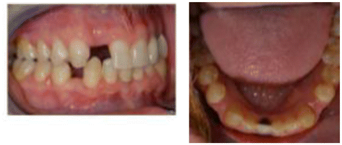
Figure 6 The patient pictured above had his snoring completely resolve after we advanced his lower anterior teeth and opened spaces for extra bicuspid implants. No recession has occurred more than 10 years after the treatment was completed.

Figure 7 Patient above, in A, was given a removable appliance to advance lower anterior teeth in a Class II case. Spaces were created between lower bicuspid teeth as a consequence of this movement, as shown in B. Temporary anchorage devices were placed bilaterally to allow for all posterior teeth to be brought forward to close the space that had been created. Effectively, the entire lower dentition was advanced over the time in treatment. The airway improved in his case as shown below (Figure ).
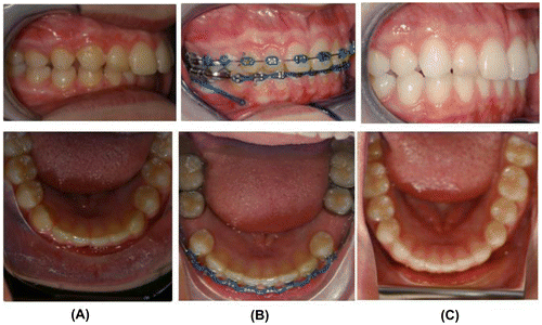
Figure 7 Patient above, in A, was given a removable appliance to advance lower anterior teeth in a Class II case. Spaces were created between lower bicuspid teeth as a consequence of this movement, as shown in B. Temporary anchorage devices were placed bilaterally to allow for all posterior teeth to be brought forward to close the space that had been created. Effectively, the entire lower dentition was advanced over the time in treatment. The airway improved in his case as shown below (Figure 8).

Figure 8 Airway improvement in a patient where lower incisors were advanced, spaces were created between lower bicuspid teeth, and posterior teeth were brought forward with TADS to close the spaces that had been created.
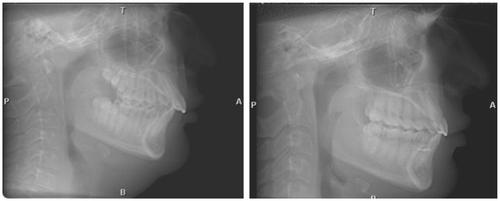
Figure 8 Airway improvement in a patient where lower incisors were advanced, spaces were created between lower bicuspid teeth, and posterior teeth were brought forward with TADS to close the spaces that had been created.

Figure 9 Patient above is a five-year old with Pierre Robin Sequence, suffering from OSA. Orthotropic® treatment to advance both jaws resulted in complete elimination of OSA.
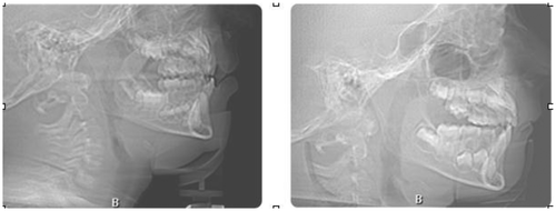
Figure 9 Patient above is a five-year old with Pierre Robin Sequence, suffering from OSA. Orthotropic® treatment to advance both jaws resulted in complete elimination of OSA.

Figure 10 This patient presented with headaches and closing loops to close the spaces remaining distal to the cuspids. After cutting the closing loops, the spaces opened slightly, relieving her headaches. B shows the space three weeks after cutting the closing loops. C shows the even larger space six weeks after cutting the closing loops.
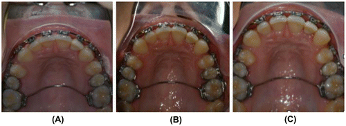
Figure 10 This patient presented with headaches and closing loops to close the spaces remaining distal to the cuspids. After cutting the closing loops, the spaces opened slightly, relieving her headaches. B shows the space three weeks after cutting the closing loops. C shows the even larger space six weeks after cutting the closing loops.