Figures & data
Table I. Clinical and demographic data in cirrhotic patients randomized to group A (synbiotic treatment) and group B (placebo treatment) and healthy controls.
Table II. Oligonucleotide primers used for real-time PCR analysis.
Figure 1. Viable faecal counts of Lactobacillus species measured pre- and post-treatment with synbiotics (left) or placebo (right). Synbiotic, but not placebo, treatment was associated with a significant increase in viable faecal counts of Lactobacillus species.
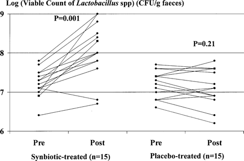
Figure 2. Indocyanine green (ICG) clearance, expressed as the percentage plasma retention rate 15 min after a test dose of ICG of 0.5 mg/kg lean body weight (ICGR15), measured pre- and post-treatment with synbiotics (left) or placebo (right). Synbiotic, but not placebo, treatment was associated with a significant improvement in ICGR15.
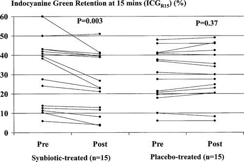
Figure 3. Serum concentrations of bilirubin (a) and albumin (b) and prothrombin time, as expressed by the international normalized ratio (INR) (c), in initially Child-Pugh class B and C patients (n=9) pre- and post-synbiotic treatment. Post-treatment values of each parameter were increased significantly compared with corresponding baseline values.
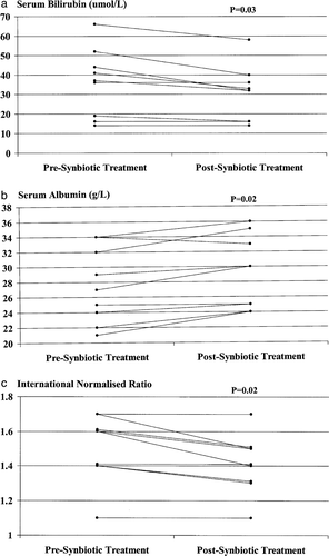
Figure 4. Whole blood TNF-α mRNA levels pre- and post-synbiotic treatment. Post-treatment values were significantly increased compared with corresponding baseline levels. *Ratio to maximum value resulting from in vitro stimulation of PBMCs by endotoxin (10 µg/ml for 20 h).
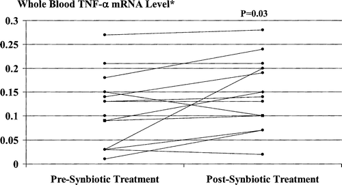
Figure 5. Whole blood IL-6 mRNA levels pre- and post-synbiotic treatment. Post-treatment values were significantly increased compared with corresponding baseline levels. *Ratio to maximum value resulting from in vitro stimulation of PBMCs by endotoxin (10 µg/ml for 20 h).
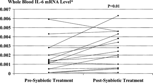
Figure 6. Serum sTNFRI levels pre- and post-synbiotic treatment. Post-treatment values were significantly increased compared with corresponding baseline levels.
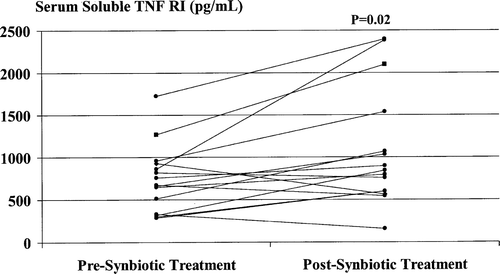
Figure 7. Serum sTNFRII levels pre- and post-synbiotic treatment. Post-treatment values were significantly increased compared with corresponding baseline levels.
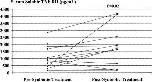
Table III. Correlations between degrees of improvement in ICGR15 and other study parameters, expressed as percentage increase or decrease compared to baseline values, in synbiotic-treated patients.
Table IV. Correlations between circulating cytokine levels in patients randomized to synbiotic treatment.