Figures & data
Figure 1. Geological information of the fossil locality at Daohugou Village. Reproduced from Han et al. (Citation2016). (a) Geographical position of Daohugou Village, Ningcheng, Inner Mongolia in China (119°14′40″E, 41°19′25″N). The rectangular region in the main map is shown in detail in the inset, in which the black triangle represents Daohugou Village and the black dots represent local cities, and (b) geological section of the Jiulongshan Formation near Daohugou Village. Layer 3 is the major fossil yielding layer. (1) gneiss; (2) tuffaceous grand conglomerate; (3) tuffaceous conglomerate; (4) tuffaceous siltstone; (5) tuffaceous mudstone; (6) tuffaceous shale; (7) volcanic breccia; and (8) fossil locality.
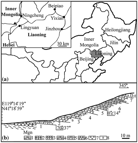
Figure 2. Yuhania daohugouensis gen. et sp. nov, and its details. Light microscopy. (a) The fossil embedded in yellowish tuffaceous siltstone. Some of the labeled regions are shown in later Figures. and 12–13 are six aggregate fruits, 14–15 are immature flowers, 5 is an associated lichen (Daohugouthallus ciliiferus (Wang et al. Citation2010a)), 6–10 are leaves, and 16 is a lateral bud. Bar = 2 cm, (b) details of the leaf marked as 7 in Figure (a), with parallel veins and entire margin. Bar = 1 mm, (c) a leaf preserved as compression to the left and as impression to the right. Bar = 1 mm, (d) detailed view of the leaf marked as 8 in Figure (a), with entire margin, alternating veins and stomata zones. Bar = 1 mm, (e) stem with longitudinal ridges, partially embedded in the sediments. Bar = 1 mm, (f) detailed view of the region as 15 in Figure (a), showing an immature flower (asterisk) in leaf (l) axil. Bar = 5 mm, (g) detailed view of the immature flower in leaf (l) axil in Figure (f). Bar = 1 mm, and (h) the aggregate fruit marked as 1 and 12 in Figure (a). Note the pedicel connected (arrow) to the stem. Bar = 2 mm.
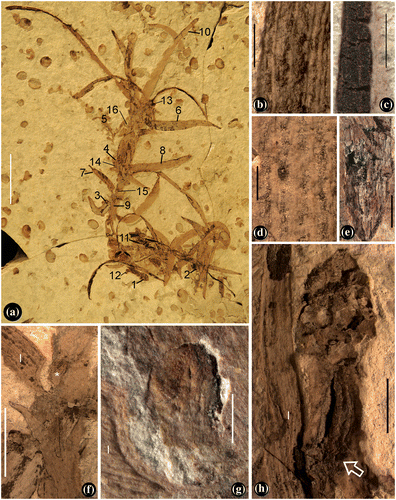
Figure 3. Leaves and their details. SEM. (a) Abaxial view of leaf tip marked as 10 in Figure (a), showing the entire leaf margin and parallel veins. Bar = 1 mm, (b) leaf tip with papillae, enlarged from the white rectangle region in Figure (a). Bar = 0.1 mm, (c) leaf texture transitional from regular (below the line) to chaotic (above the line), enlarged from the black rectangle in Figure (a). Bar = 0.2 mm, (d) an adaxial view of a leaf, showing longitudinal epidermal cells and entire leaf margin (arrow). Bar = 0.1 mm, (e) an abaxial view of the leaf in Figure (d), showing well-defined alternating vein and intervein (stomata, arrow) zones. Bar = 0.2 mm, (f) leaf (l) clasping and diverging from the stem (s) with horizontal wrinkles. Note the leaf texture changes from the horizontal to longitudinal from the bottom up. Bar = 0.2 mm, (g) detailed view of the stomata arrowed in Figure (e). Bar = 0.1 mm, (h) a leaf with elongate epidermal cells (upper-left) and mesophyll aerenchyma. Bar = 50 μm, (i) a leaf in its earliest developmental stage, fringed with dentate protrusions. Bar = 0. 1 mm, and (j) leaf probably damaged by insect (arrow). Bar = 0.5 mm.
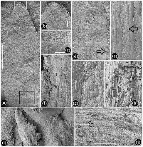
Figure 4. Flower and aggregate fruits of Yuhania. SEM. (a) The immature flower in Figure (g), with a stout pedicel and spherical receptacle. Bar = 0.5 mm, (b) detailed view of the rectangle in Figure (a), showing outlines of the carpels helically arranged. Bar = 20 μm, (c) the sac-like carpel marked as C in Figure (a). Bar = 10 μm, (d) SEM view of the aggregate fruit in Figure (h), with helically arranged fruitlets. Bar = 0.5 mm, (e) one of the fruitlets from the aggregate fruit in Figure (d), with its seed exposed. Bar = 0.2 mm, (f) detailed view of the distal portion of the fruitlet in Figure (e). Note the cuspidate tip (black arrow), the greatest width near the distal of the fruitlet, and a bract (white arrow). Bar = 0.1 mm, (g) detailed view of the proximal part of the fruitlet in Figure (e), showing the broken fruitlet wall (arrows) and exposed seed (s) in the fruitlet. Bar = 50 μm, (h) rounded tip of a bract, note the longitudinal texture in the middle. Bar = 0.1 mm, (i) SEM view of the aggregate fruit marked as 2 in Figure (a) and shown in Figure S4(e) and (f). Bar = 0.5 mm, (j) a young ‘carpel’ with a broken tip (black arrow), wide base, and a bract in the background (white arrow). Note the empty space in the ‘carpel.’ Bar = 0.2 mm, (k) a young fruitlet with an extended terminus (arrow). Bar = 0.5 mm, (l) detailed view of young fruitlet shown in Figure (j), showing outline of a possible ovule (white line). Bar = 50 μm, and (m) detailed view of young fruitlet shown in Figure (j), showing ovarian wall (ow) fused (arrow) to the floral axis (fa). Bar = 0.1 mm.
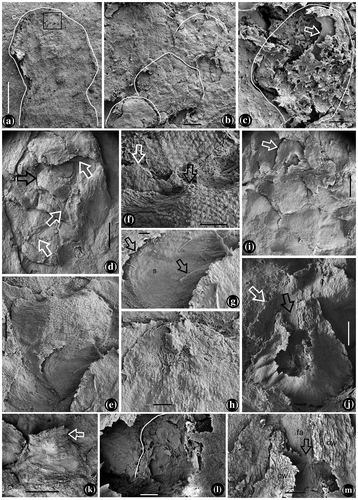
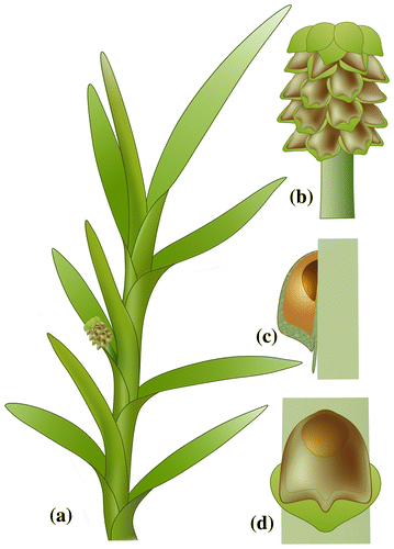
Figure 5. Reconstruction of Yuhania. (a) Shoot with leaves and aggregate fruit, (b) pedicellate aggregate fruit, (c) longitudinal section of a carpel/fruitlet, showing an ovule/seed inserted on floral axis and enclosed in ovary, and (d) surface view of a carpel/fruitlet, showing an ovule/seed inserted on floral axis and enclosed in ovary.