Figures & data
Figure 1. Photographs and patterns of the degree d and angle α of linear polarisation of the sticky real brown, painted black and painted white vertical maple tree trunk pieces measured with imaging polarimetry in the green (550 nm) spectral range in field experiment 3. The polarimeter’s optical axis was horizontal. The trunks were in the shade of trees and thus illuminated by light from the clear sky and the surrounding vegetation. In the α-pattern the white bars show the local direction of polarisation of the painted bark
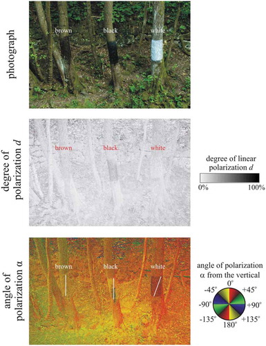
Figure 2. Photographs and patterns of the degree d and angle α of linear polarisation of the sticky region of two fallen horizontal brown maple tree trunks (a and b are the same but seen from two different viewing directions) overarching the mountain creek used in experiment 4 measured with imaging polarimetry in the green (550 nm) spectral range. The tilt angle of the polarimeter’s optical axis was −35° from the horizontal. In the α-pattern the white bars show the local direction of polarisation of the sticky bark region. In case a and b the scene was in the shade of trees and illuminated by light from the clear sky and the surrounding vegetation. In case c, beyond skylight, the scene was illuminated from the left side by direct light of the setting sun

Figure 3. Photographs and patterns of the degree d and angle α of linear polarisation of the colourless insect-monitoring glue used in experiments 2–4 (1), the yellowish cooking oil used in experiment 1 (2), a yellowish Canada balsam (3), and a brown pine resin dissolved in isopropyl alcohol (4) measured with imaging polarimetry in the green (550 nm) spectral range under shady (left column) and sunlit (right column, when the sun shone frontwise) conditions when the Petri dishes containing the sticky liquids were on a black, brown and white substrate. The tilt angle of the polarimeter’s optical axis was −65° from the horizontal. In the α-patterns the white bars show the local direction of polarisation of the liquid surface
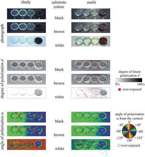
Table 1. Number of the chironomid flies trapped by the black, brown, light brown and white trays padded with bark and filled with cooking oil in experiment 1. χ2 test of homogeneity yielded significant differences among the sums (χ2 = 1200.7, df = 3, p < 0.0001)
Table 2. Numbers of trapped aquatic insects on the three vertical (real brown, painted black, painted white) and one horizontal (real brown) sticky trunk surfaces. Females and males are indicated by ‘f’ and ‘m’ in brackets, respectively. In the case of mayflies (Ephemeroptera), ‘s’ and ‘i’ denote subimago and imago. χ2 test of homogeneity yieldedsignificant differences among the sums of specimens (χ2 = 6.3, df = 2, p < 0.05)
Figure 4. Mayflies trapped by the real brown (a–b), painted black (c–d) and painted white (e–f) vertical sticky barks in experiment 3. (a, c) Female Ephemera danica subimagoes. (b–d) Female Rhithrogena semicolorata subimagoes. (e) Male Ephemera danica subimago. (f) Male Epeorus silvicola imago
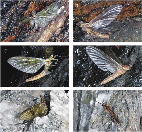
Figure 5. Mayflies trapped by the horizontal sticky bark in experiment 4. (a–b) Male and female Ephemera danica imagoes. (c) Male Epeorus silvicola. (d) Female R. semicolorata with her egg-batch
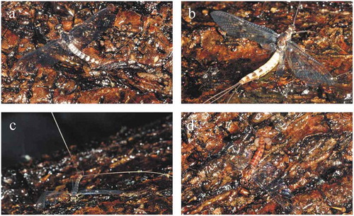
Figure 6. (a–f) Aquatic insects trapped by the painted black vertical sticky bark in experiment 3. (a) Beautiful demoiselle (Calopteryx virgo) female. (b) Female C. virgo exhausted during emergence. (c–d) Stoneflies (Plecoptera). (c) Common forestfly (Nemoura cinerea). (d) Sallfly (Chloroperla sp.). (e) Caddisfly (Rhyacophilidae). (f) Crane fly (Tipulidae)
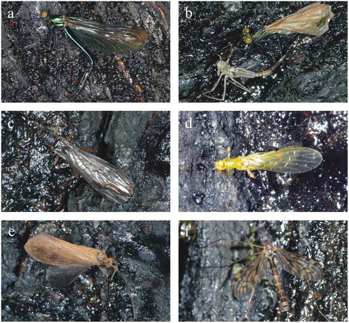
Figure 7. Aquatic insects trapped by the real brown vertical sticky bark in experiment 3. (a) Beautiful demoiselle (Calopteryx virgo) male. (b) Male C. virgo exhausted during emergence. (c–d) Stoneflies (Plecoptera). (c) Common forestfly (Nemoura cinerea). (d) Nymphal skin of Perla abdominalis. (e) Alderfly (Sialis sp.). (f–g) Caddisflies. (f) Philopotamus sp. (g) Hydropsychidae. (h) Crane fly (Tipulidae)
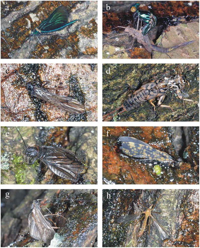
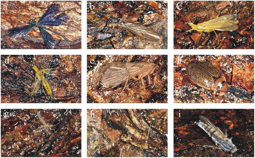
Figure 8. Aquatic insects trapped by the painted white vertical sticky bark in experiment 3. (a) Nymphal skin of Calopteryx virgo. (b–c) Stoneflies (Plecoptera). (b) Common forestfly (Nemoura cinerea). (c) Isoperla tripartita. (d–e) Caddisflies (Trichoptera). (d) Rhyacophilidae. (e) Limnephilidae. (f) Crane fly (Tipulidae)
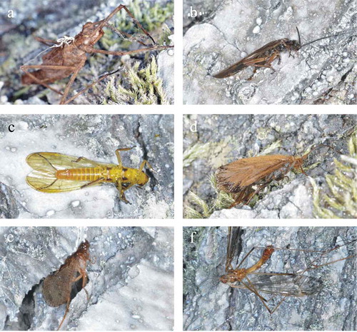
Figure 9. Aquatic insects trapped by the real brown horizontal sticky bark in experiment 4. (a) Beautiful demoiselle (Calopteryx virgo) male. (b–d) Stoneflies (Plecoptera). (b) Common forestfly (Nemoura cinerea). (c) Isoperla tripartita. (d) Sallfly (Chloroperla sp.). (e) Caddisfly (Limnephilidae). (f) Diving beetle (Colymbetes fuscus). (g) Female non-biting midge (Chironomidae). (f) Crane fly (Tipulidae). (h) Horsefly (Tabanidae)